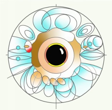
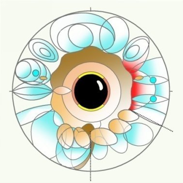
Francesco Puerari
The basics of Iridology
Maps
Summary
About the author
Francesco Puerari MD has worked in an Italian General Hospitals Anesthesia and Intensive Care Unit for 34 years. He has earned several postgraduate specializations (Anesthesia and Intensive Care, Dietetics and Nutrition, Medical Toxicology, Neurology) at the University of Pavia. All along his professional life, he has also attended complementary medicine resources (Iridology and Homeopathy) thus enriching his medical approach.
Work plan
This is the second of three textbooks on Iridology, a discipline focused on analyzing the information given by the colored part of the eye called iris. It will focus on a detailed description of the organs projections in the iris (Maps).
The first textbook has described the morphological variants of the iris (Iris patterns).
The third textbook will be dedicated to the signs of unbalance collected in the iris (Markings).
Acknowledgements
I want to thank the masters of Iridology E. Ratti, F. Minisini, J. Karl, W. Hauser, R. Stolz, A. A. Sartorelli, L. Birello and the Italian Iridology Association (ASSIRI).
Copyright
Copyright 2014 by Francesco Puerari. All right reserved. No part of this book may be reproduced or transmitted in any form or by any means, graphic, electronic, or mechanical, including photocopying, recording, taping or by any information storage or retrieval system without the written permission of the author/publisher.
Published by: Francesco Puerari
Cover, tables and pictures by: Francesco Puerari
Editing by: Paolo Folzini/Francesco Puerari
ISBN: 978-88-940272-0-4
www.irispatterns.it
Iridology a definition
Iridology studies the colored portion of the eye named iris.
The iris is a highly innervated organ that is stimulated both by the external environment and by the body.
The structure of the iris mirrors the individual constitution; illnesses, harmful habits and aging can alter it.
The iris analysis completes medical practice by supplying data on constitution, nervous response, damages caused by aging, illnesses and familiarity.
Iridology is a discipline that enriches traditional investigations. It collects signs. It does not provide diagnosis.
This book is an information source only. It does not provide advice for self-diagnosis or self-prescription and treatment.
MAPS OF THE IRIS
The connections and interactions between internal organs and bodys surface are utilized in several fields of health care: Traditional Chinese Acupuncture, Reflexology, Kinesiology
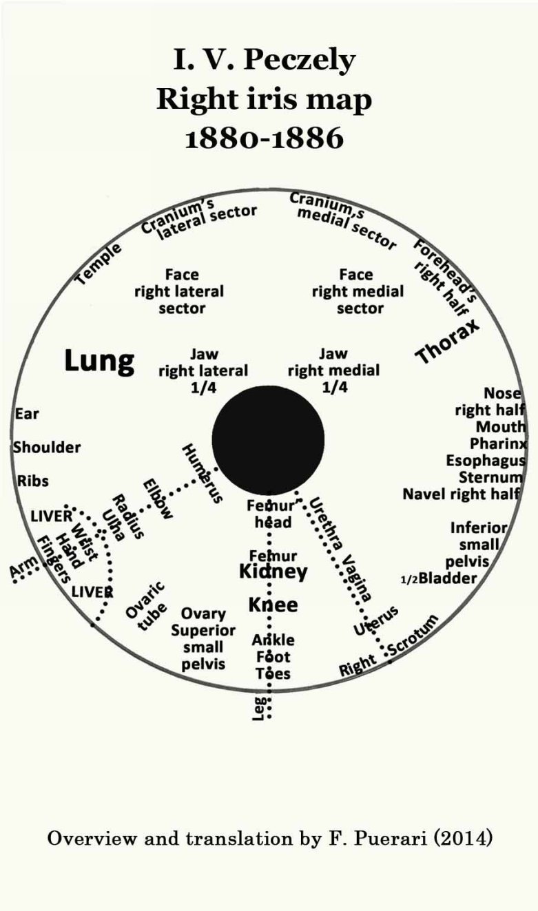
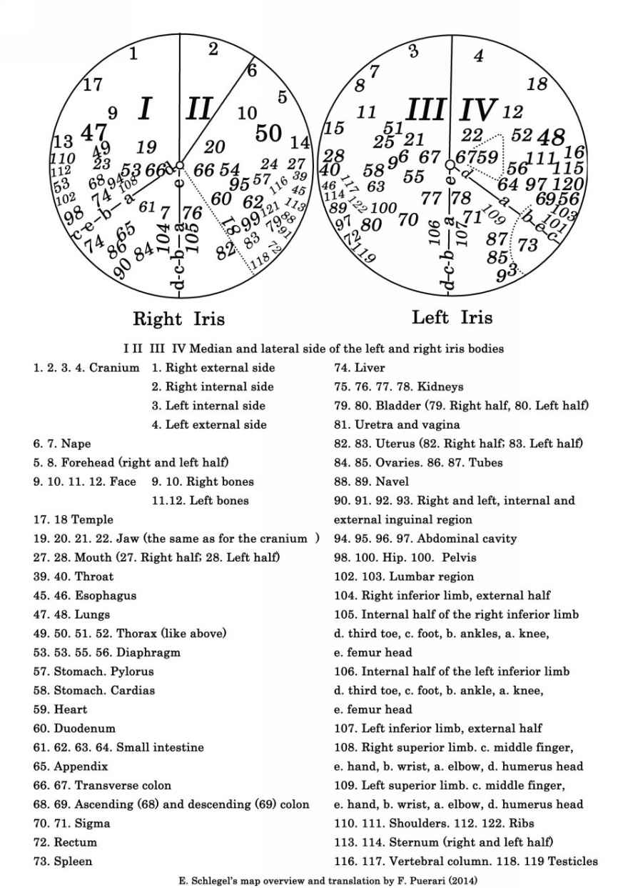
The Hungarian physician I.V. Peczely (1826-1911) realized that this approach could also be applied to the iris surface. In 1871 Peczely started to divulge his discoveries in conferences and publications. In 1880 he published his map of human internal organ projections in the iris (Entdeckungen auf dem Gebiete der Natur und Heilkunde, Budapest, 1880). The first physician who recognized the scientific reliability of his discoveries was Emil Schlegel who published, in 1886, a re-elaboration of Peczely original map thus acknowledging his fundamental contributions (Die Augendiagnose der Dr. Ignacz Von Peczely, Tubingen, 1886).
Peczely and Schlegel are considered as the founders of modern iridology. The version of their maps and diagrams provided in this e-book is a re-elaboration and translation of different sources which aims at respecting the original ones as much as possible.
I. PECZEL Y S AND SHLEGEL MAPS
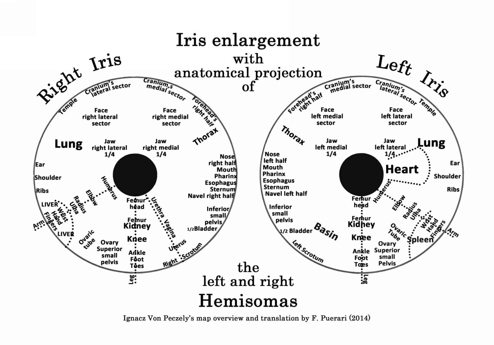
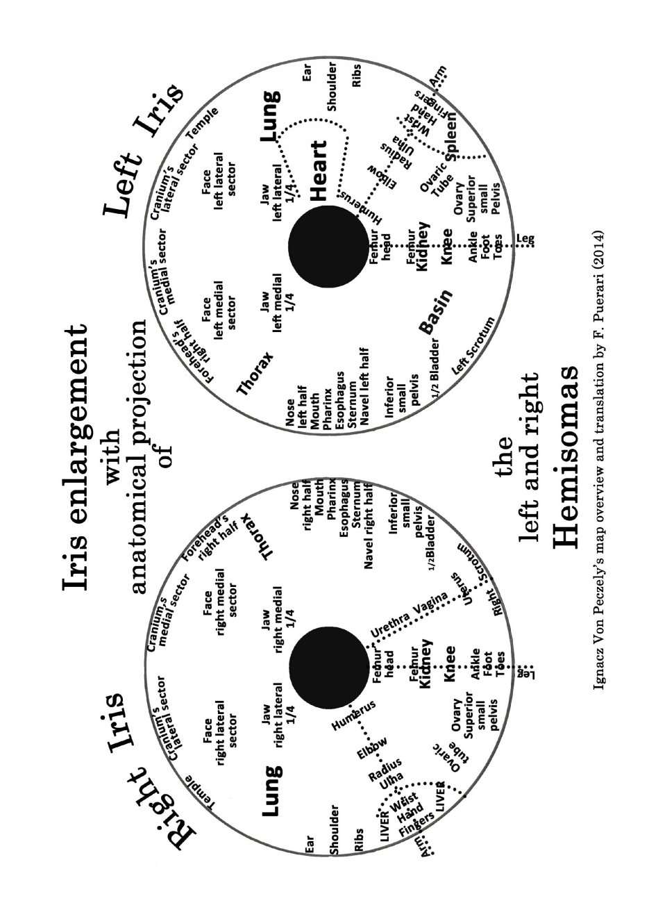
Peczely map shows the organ names in their corresponding projection areas. Schlegels map numbers the organs projection points on the iris chart and provides their reading in an added table. Emil Schlegel also provided several iris diagrams resulting from his clinical findings. The maps suggested below and in the next pages are a translated compendium of Peczely and Schlegels works, aimed at grasping the meaning of the two scholars research.
PECKZELY MAP TRANSLATION
(F. PYERARI 2014)
E. SCHLEGELS MAP TRANSLATION
(F. Puerari 2014)
E. SCHLEGELS CLINICAL REPORT MAP (1886)
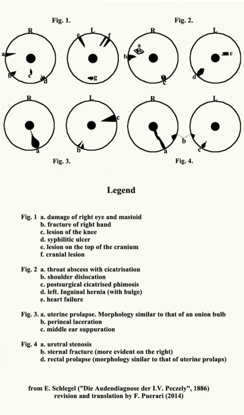
In the decades following the publication of these maps, Nils Liljequist (1851-1936) realized that quinine, a drug commonly prescribed at that time, could often saturate the iris with toxic pigments and that bone fractures were often cause of changes in the iris. Liljequist published his first works, rich in maps and tables, in 1893. Since then, many different iris maps have been published and many iridological schools of thought are born.
The maps elaborated by the German iridological school are widely shared (W. Hauser, J. Karl, R. Stolz "Die praktische Iris diagnostik", Koln-Heimsheim, 1986; Informatonen aus Struktur und Farbe Felke Institut Heimsheim Germany, 2001). However, there are numerous other maps that are considered authoritative. The most used ones being the maps of J. Deck, S. J. Rizzi, L. Berdonces, B. Jensen (J. Deck Differenzierung der iris zeichen Karlruhe, 1965; S. Rizzi, "Iridology, the future diagnostic method", I. Laches 1983; J. L. Berdonces, "Basic Manual of Iridology", Integral Ediciones, Oasis, Barcelona 1990; B. Jensen, Iridology, Escondido, 1982). Finally, it is also worth mentioning the chart recently developed by E. Ratti and J. Karl (Iridologie Bildatlas mit Erlauterung, Autonome Provinz Bozen Sudtirol, 2011).
Circular and sectorial map
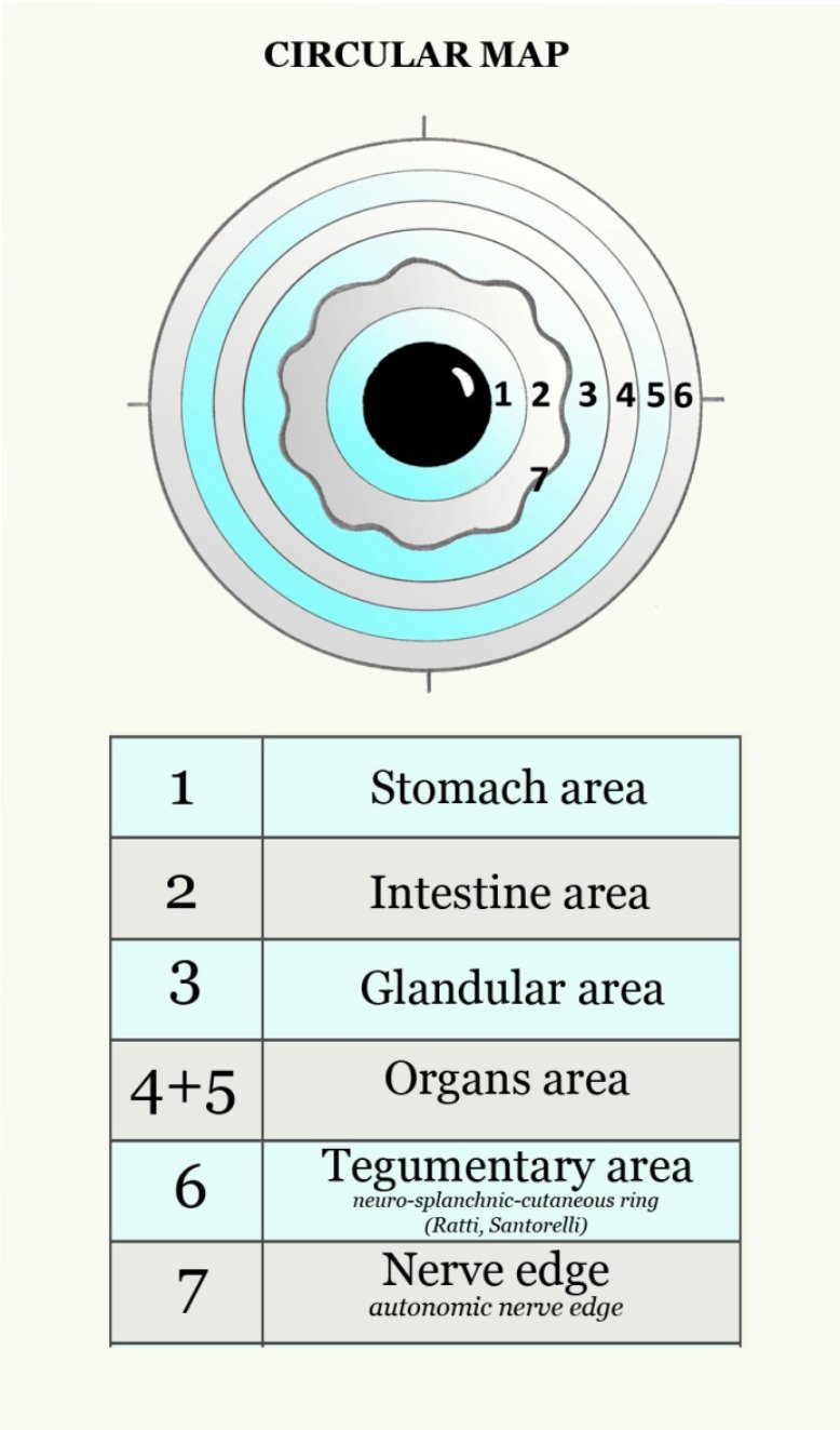
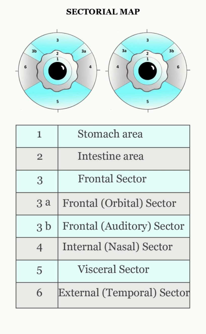
The iris subdivision in concentric circles (circular map) or in radial sectors (sectorial map), simplifies iris analysis and allows for a correct localization of the points of interest.
Circular map. The circular subdivision divides the iris into six concentric circles that coincide with as many areas and functions of the human body.
Sectorial map. The eight sectors of the sectorial subdivision coincide with the major apparatuses.
OVERLAPPING
The maps submitted in this e-book are a compendium of the most shared knowledge.
Next page


