Evan H. Black (editor) - Smith and nesis Ophthalmic Plastic and Reconstructive Surgery
Here you can read online Evan H. Black (editor) - Smith and nesis Ophthalmic Plastic and Reconstructive Surgery full text of the book (entire story) in english for free. Download pdf and epub, get meaning, cover and reviews about this ebook. year: 2012, publisher: Springer Nature, genre: Home and family. Description of the work, (preface) as well as reviews are available. Best literature library LitArk.com created for fans of good reading and offers a wide selection of genres:
Romance novel
Science fiction
Adventure
Detective
Science
History
Home and family
Prose
Art
Politics
Computer
Non-fiction
Religion
Business
Children
Humor
Choose a favorite category and find really read worthwhile books. Enjoy immersion in the world of imagination, feel the emotions of the characters or learn something new for yourself, make an fascinating discovery.

- Book:Smith and nesis Ophthalmic Plastic and Reconstructive Surgery
- Author:
- Publisher:Springer Nature
- Genre:
- Year:2012
- Rating:3 / 5
- Favourites:Add to favourites
- Your mark:
Smith and nesis Ophthalmic Plastic and Reconstructive Surgery: summary, description and annotation
We offer to read an annotation, description, summary or preface (depends on what the author of the book "Smith and nesis Ophthalmic Plastic and Reconstructive Surgery" wrote himself). If you haven't found the necessary information about the book — write in the comments, we will try to find it.
Smith and Nesis Ophthalmic Plastic and Reconstructive Surgery, Third Edition has taken the best of the fields classic reference text and expanded upon it, continuing its reputation as the foremost guide to the subspecialty. Every practitioner of plastic and reconstructive surgery will find useful information in this comprehensive, in-depth text, including an update on the changes and advances of the last several years and a new section on pediatric consideration. Detailed chapters on ophthalmic anatomy are also included, as well as sections on eyelid dermatology, socket surgery, and cosmetic surgery. Every consideration has been made by Dr. Nesi and his co-editors to cover every aspect important to the ophthalmic plastic surgeon.
Features:
Fully updated and revised edition of Smiths Ophthalmic Plastic and Reconstructive Surgery
Comprehensive text with detailed chapters that cover all aspects of the subject matter
More than 1700 figures and 77 chapters
Evan H. Black (editor): author's other books
Who wrote Smith and nesis Ophthalmic Plastic and Reconstructive Surgery? Find out the surname, the name of the author of the book and a list of all author's works by series.


Smith and nesis Ophthalmic Plastic and Reconstructive Surgery — read online for free the complete book (whole text) full work
Below is the text of the book, divided by pages. System saving the place of the last page read, allows you to conveniently read the book "Smith and nesis Ophthalmic Plastic and Reconstructive Surgery" online for free, without having to search again every time where you left off. Put a bookmark, and you can go to the page where you finished reading at any time.
Font size:
Interval:
Bookmark:
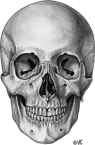
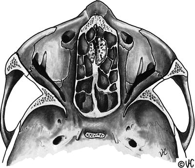
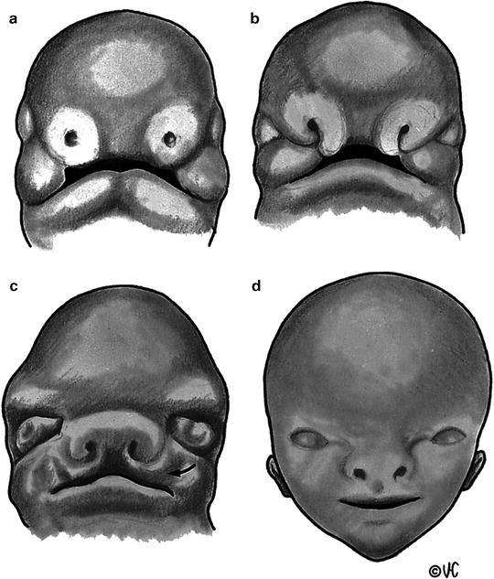
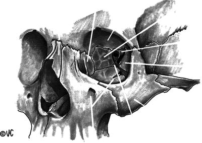
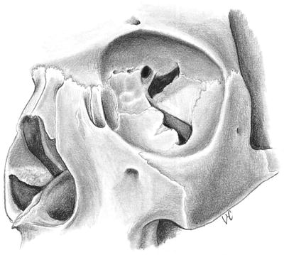
Anatomy
Font size:
Interval:
Bookmark:
Similar books «Smith and nesis Ophthalmic Plastic and Reconstructive Surgery»
Look at similar books to Smith and nesis Ophthalmic Plastic and Reconstructive Surgery. We have selected literature similar in name and meaning in the hope of providing readers with more options to find new, interesting, not yet read works.
Discussion, reviews of the book Smith and nesis Ophthalmic Plastic and Reconstructive Surgery and just readers' own opinions. Leave your comments, write what you think about the work, its meaning or the main characters. Specify what exactly you liked and what you didn't like, and why you think so.