About This Book
In the past, a lot of individual surgery procedures and histopathological investigation for skin tumors have been extensively discussed in the literature, e.g., Mohs surgery and methods of other disciplines as well. This interdisciplinary book attempts to summarize and compare the most commonly used methods in order to give the reader the most comprehensive and complete overview of the current state of the art in the field. To this end, the basic principles of various surgical methods and following histological workups of the complete 3D tumor margins will be described and extensively graphically illustrated to allow a better understanding. Furthermore, also more complex treatments of larger tumors will be discussed and comprehensively illustrated.
It should be noted that all methods described here are exchangeable and can be freely combined much to the liking of the executing surgeon and pathologist. Clinically, there are only minor differences between most of the described procedures in this book. This will allow the reader to come up with his or her own preferred choice of surgery and histological control which will help to assess and streamline the currently used workflow.
Not only surgical skills and histological preparations and evaluations are important for a successful and efficient treatment of various malignant skin tumors, also a good knowledge of local patterns of infiltration is required to achieve complete excision. All tumors may show an irregular, often asymmetric, mainly horizontal subclinical infiltration with a lateral extent of zero to several centimeters within the epidermis, dermis, and upper subcutaneous tissue. When in-depth infiltrations are present, they are often asymmetric as well and hard to determine before excision. Up to now, these tumor outgrowths are only detectable by a histological evaluation. 3D histology is highly sensitive and can detect even very small tumor infiltrations; therefore, very narrow margins can be taken for excision. This helps sparing healthy skin as only a subsequently targeted re-excision will be necessary to remove all of the asymmetric tumor infiltrations. Therefore, the resulting defect will be as small as possible and as large as necessary. The resulting shape of the defect can be adapted to the requirement of the rules for aesthetic surgical defect closure.
Also the organization of the workflow, the scheme of documentation used, and the available infrastructure greatly influence the efficiency and accuracy of the procedure. This directly improves the patients welfare, as less and more targeted surgical procedures with a high reproducibility can be performed. Hence, the importance of workflow organization and documentation cannot be underestimated. Consequently, an entire chapter of this book is dedicated to this critical aspect of skin tumor treatment.
The main emphasis of this book lays on a thorough and comprehensive illustration of the methods. Therefore, the illustrations should be self-explaining and may not require any further explanations. However, every figure is accompanied by comprehensive description in order to give the reader the best and most complete introduction to all discussed methods.
What Is 3D Histology?
3D histology provides a complete 3D representation of the margins of a tumor specimen on histological slides (Table ).
Table 1.1
Definition of 3D histology
1.Complete 3D margins of a tumor specimen in histological slides |
2.No diagnostic gaps |
3.The highest sensitivity to make tumor outgrowths topographical visible |
4.Tumor-positive areas are removed by step-by-step re-excisions until the margins are clear |
5.Saving healthy tissue. Minimal invasive surgery |
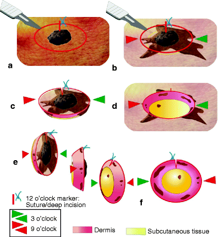
Figure 1.1
Tumor with subclinical infiltrations. After excision and flipping over, the 3D margin outside of the specimen can be seen. ( a ) The clinically visible tumor with excisional margins. A suture or deep incision at 12 oclock relative to the patients top of the head serves as a landmark. Tumor resection line in red . ( b ) Subclinically, normally invisible tumor infiltrations are shown in brown for purposes of illustration. ( c ) The excised tumor with its surrounding tissue and 3D borders. ( d ) The defect after excision. ( e ) Illustration of the bowl-shaped tumor specimen as it is flipped over to see its reverse side. Colored arrowheads serve for orientation ( green arrowheads 3 oclock, red arrows 9 oclock). ( f ) The complete 3D view of the excisional margins from beneath with subclinical, infiltration sites in brown . These infiltrations only become visible after histological staining
It should be mentioned that in the figures of this book, tumor outgrowths which might have been missed in the first excision are highlighted as brown spots at the margins of the tumor specimens and in the resulting defect for better visualization of the reader. These outgrowths are clinically invisible and can only be seen histologically after staining with H&E. They are intended to show the complexities and variability of tumor outgrowths and should help to understand the importance of 3D histology.
The basic principle of 3D histology is the conversion from the 3D structure of the tumor-specimen margins into a 2D view of histological sections to detect tumor infiltrations. The 3D margins are flattened with their outside down and then cut into pieces, suitable for histopathological procedures of cryo- or paraffin sections (see ).
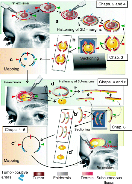
Tumor-infiltrated areas can then be removed step by step by targeted re-excisions until the margins are clear, thus sparing healthy tissue. This minimal invasive surgery ensures that the defect will be as small as possible especially in difficult localizations. The next figure gives an overview over the workflow using 3D histology and the corresponding chapters of this book in which extensive explanations will be given (Fig. ).
Figure 1.2
Short workflow of using 3D histology. Tumor excision, flattening of the 3D margins, sectioning, mapping, and re-excisions. ( a ) Excision of the tumor of Fig. ) and ( d ) second and third re-excision

