1.1 Introduction
A wide variety of neonatal and childhood dermatologic conditions can be emergencies. These may arise from genetic disorders, including those causing skin structural protein and keratin dysfunction, vascular birthmarks with systemic associations, autoimmune disorders, and infections. If left untreated, these conditions can lead to systemic effects or may increase the risk of developing secondary complications, potentially resulting in death. It is therefore important that these entities be rapidly identified and addressed. The following pages present a limited collection of some of the most important emergency situations.
1.2 Neonatal Emergencies
There are broad sets of neonatal dermatologic conditions that are true emergencies. Vesicles, pustules, and denuded skin at birth or in the neonatal period may be localized lesions without consequence, such as sucking blisters, or signs of a life-threatening genetic mechanobullous disorder such as epidermolysis bullosa. Herpes simplex virus (HSV) and opportunistic infections can be dermatologic emergencies, requiring prompt recognition, diagnosis, and initiation of therapy. Vascular tumors, including port-wine stains (PWS) and hemangiomas, may require urgent management including diagnostic workup, referral for specialty evaluation, and/or early introduction of therapy. The conditions discussed below are only a select subset of neonatal dermatologic issues that may be considered emergencies. Other resources more extensively discuss other neonatal dermatologic conditions (e.g., Neonatal Dermatology , 2nd Edition. Eichenfield, Frieden and Esterly, Saunders/Elsevier).
1.3 Epidermolysis Bullosa
Epidermolysis bullosa (EB) is a group of congenital disorders characterized by the profound susceptibility of skin and mucosa to separate from underlying tissues following mechanical trauma. EB generally presents at birth or in the first few weeks of life with denuded skin, vesicles, or bullae. Milder forms may present later in life. EB is caused by mutations in structural proteins in the skin, and is generally categorized by the level of blistering and the target proteins: EB simplex (EBS) (epidermolytic), junctional EB (JEB) (lysis in the lamina lucida, or lucidolytic), dystrophic (dermolytic), and Kindler syndrome (mixed blistering levels) [].
EB has an incidence of 1 in every 50,000 births, consisting mostly of the milder EBS type []. Oral erosions are most commonly found in JEB and DEB, and are rarely found in EBS. Extensive blistering can lead to fluid and electrolyte abnormalities, temperature dysregulation, as well as sepsis; supportive measures are essential in the treatment plan.
The differential diagnosis of erosions, blisters, or denuded skin includes infections (e.g., HSV) and other blistering diseases. To diagnose EB, a biopsy should be taken from unaffected skin. The area to be biopsied may be rubbed for 1 min, generally with a pencil eraser, to induce cutaneous blistering; after a 510-min wait, a shave, or punch biopsy is taken []. Evaluation can include light microscopy, immunofluorescence antigen mapping for absent or attenuated antigens diagnostic for the EB subtype, or transmission electron microscopy, which may visualize the ultrastructural level exhibiting the plane of separation. Specific genetic testing may be utilized as well.
1.3.1 Epidermolysis Bullosa Simplex
EBS, the mildest and most common type of EB, is characterized by the lysis of basal keratinocytes above the basement membrane zone [].
1.3.2 Junctional Epidermolysis Bullosa
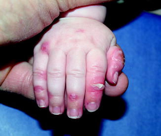
JEB (Fig. ].
Fig. 1.1
Junctional epidermolysis bullosa. Erosions, blisters, and denudation on the hands
1.3.3 Dystrophic Epidermolysis Bullosa
DEB is characterized by blistering below the lamina densa of the basement membrane zone, causing lysis of the dermis [].
Emergent evaluation of a neonate with possible EB includes skin biopsy and institution of aggressive supportive measures, including excellent skin care. Fluids and electrolytes must be closely monitored, and the temperature must be kept moderate since heat can induce blistering. Care must be taken to minimize new blisters and erosions through gentle handling and the use of nonadhesive bandages especially for the first layer of bandaging. The skin care regimen must promote wound healing through daily bathing and vigilant inspection of blisters. A topical or systemic antimicrobial can be used to prevent infection, though use must be balanced by the potential for selection of resistant microbes. Topical antibiotics may be rotated and the use of such products should be discontinued when the wound is clean []. Nonsteroidal anti-inflammatory drugs or opiates can be administered to decrease pain during bandaging and other procedures. Intact blisters must be incised on the dependent side using a sterile needle to prevent lesion extension. It is important to properly diagnose the EB type and subtype, as well as counsel and educate parents on prognosis and future therapy. Referral for genetic counseling is appropriate. In-hospital care may be appropriate until the infant is feeding well and gaining weight with stable skin involvement, and the family has adequate training in skin care management.
1.4 Disorders of Keratinization (Ichthyosis)
There are several ichthyotic conditions that manifest during the neonatal period as either collodion baby or scaling erythroderma. These disorders encompass a wide range of genetic conditions with molecular defects affecting the epidermis []. For most ichthyotic conditions, early recognition, therapy, and monitoring will be required to prevent infection or fluid and electrolyte imbalance.
1.4.1 Collodion Baby
The collodion infant presents at birth encased in a shiny, thickened, variably erythematous, cellophane-like membrane [].

