Marc G. Jeschke , Lars-Peter Kamolz and Shahriar Shahrokhi (eds.) Burn Care and Treatment 2013 A Practical Guide 10.1007/978-3-7091-1133-8_1 Springer-Verlag Wien 2013
1. Initial Assessment, Resuscitation, Wound Evaluation and Early Care
Shahriar Shahrokhi 1
(1)
Division of Plastic and Reconstructive Surgery, Ross Tilley Burn Centre, Sunnybrook Health Sciences Centre, 2075 Bayview Ave, Suite D716, Toronto, ON, M4N 3M5, Canada
Abstract
The initial assessment and management of a burn patient begins with prehospital care. There is a great need for efficient and accurate assessment, transportation, and emergency care for these patients in order to improve their overall outcome. Once the initial evaluation has been completed, the transportation to the appropriate care facility is of outmost importance. At this juncture, it is imperative that the patient is transported to facility with the capacity to provide care for the thermally injured patient; however, at times patients would need to be transported to the nearest care facility for stabilization (i.e., airway control, establishment of IV access).
1.1 Initial Assessment and Emergency Treatment
The initial assessment and management of a burn patient begins with prehospital care. There is a great need for efficient and accurate assessment, transportation, and emergency care for these patients in order to improve their overall outcome. Once the initial evaluation has been completed, the transportation to the appropriate care facility is of outmost importance. At this juncture, it is imperative that the patient is transported to facility with the capacity to provide care for the thermally injured patient; however, at times patients would need to be transported to the nearest care facility for stabilization (i.e., airway control, establishment of IV access).
Once in the emergency room, the assessment as with any trauma patient is composed of primary and secondary surveys (Box ). As part of the primary survey, the establishment of a secure airway is paramount. An expert in airway management should accomplish this as these patients can rapidly deteriorate from airway edema.
Once this initial assessment is complete, the disposition of the patient will be determined by the ABA criteria for burn unit referral [).
Table 1.1
ABA criteria for transfer to a burn unita
1. Partial-thickness burns greater than 10 % total body surface area (TBSA) |
2. Burns that involve the face, hands, feet, genitalia, perineum, or major joints |
3. Third-degree burns in any age group |
4. Electrical burns, including lightning injury |
5. Chemical burns |
6. Inhalation injury |
7. Burn injury in patients with preexisting medical disorders that could complicate management, prolong recovery, or affect mortality |
8. Any patient with burns and concomitant trauma (such as fractures) in which the burn injury poses the greatest risk of morbidity or mortality. In such cases, if the trauma poses the greater immediate risk, the patient may be initially stabilized in a trauma center before being transferred to a burn unit. Physician judgment will be necessary in such situations and should be in concert with the regional medical control plan and triage protocols |
9. Burned children in hospitals without qualified personnel or equipment for the care of children |
10. Burn injury in patients who will require special social, emotional, or rehabilitative intervention |
aFrom Ref. []
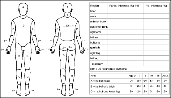
In determining the %TBSA (% total body surface area) burn, the rule of 9 s can be used; however, it is not as accurate as the Lund and Browder chart (Fig. ) which further subdivides the body for a more accurate calculation. First-degree burns are not included.
Fig. 1.1
Lund and Browder chart for calculating %TBSA burn
Assessment of burn depth can be precarious even for experts in the field. There are some basics principles, which can help in evaluating the burn depth (Table ). Always be aware that burns are dynamic and burn depth can progress or convert to being deeper. Therefore, reassessment is important in establishing burn depth.
Table 1.2
Typical clinical appearance of burn depth
First-degree burns | Involves only the epidermis and never blisters |
Appears as a sunburn |
Is not included in the %TBSA calculation |
Second-degree burns (dermal burns) | Superficial |
Pink, homogeneous, normal cap refill, painful, moist, intact hair follicles |
Deep |
Mottled or white, delayed or absent cap refill, dry, decreased sensation or insensate, non-intact hair follicles |
Third-degree burns | Dry, white or charred, leathery, insensate |
Given that even burn experts are only 6476 % []:
Table 1.3
Techniques used for assessment of burn deptha
Technique | Advantages | Disadvantages |
|---|
Radioactive isotopes | Radioactive phosphorus (32P) taken up by the skin | Invasive, too cumbersome, poorly reproducible |
Nonfluorescent dyes | Differentiate necrotic from living tissue on the surface | No determination of depth of necrosis; many dyes not approved for clinical use |
Fluorescent dyes | Approved for clinical use | Invasive; marks necrosis at a fixed distance in millimeters, not accounting for thickness of the skin; large variability |
Thermography | Noninvasive, fast assessment | Many false positives and false negatives based on evaporative cooling and presence of blisters; each center needs to validate its own values |
Photometry | Portable, noninvasive, fast assessment, validated against senior burn surgeons, and color palette was developed | Single-institution experience; expensive? |
Liquid crystal film | Inexpensive | Contact with tissue required, unreliable readings |
Nuclear magnetic resonance | Water content in tissue differentiates partial from full-thickness wounds | In vitro assessment only, expensive, time-consuming |
Nuclear imaging | 99mTc shows areas of deeper injury | Expensive, very time-consuming, not readily available, and invasive |
Pulse-echo ultrasound | Noninvasive, easily available | Underestimates depth of injury, operator-dependent, and requires contact with tissue |
Doppler ultrasound | Noncontact technology available, provides morphologic and flow information | Operator-dependent, not as reliable as laser Doppler |

