Part 1
Primary Burn Wound Management
Hiko Hyakusoku , Dennis P. Orgill , Luc Teot , Julian J. Pribaz and Rei Ogawa (eds.) Color Atlas of Burn Reconstructive Surgery 10.1007/978-3-642-05070-1_1 Springer-Verlag Berlin Heidelberg 2010
1. Primary Wound Management: Assessment of Acute Burns
Luc Tot 1
(1)
Montpellier University, Montpellier, France
Abstract
The burn is depicted as a traumatic lesion provoked by several possible agents (thermal, chemical, mechanical, or electrical) involving different skin layers to a certain degree. Assessment of the clinical situation is based on (1) evaluation of the total body surface of the burns, and (2) estimation of burn depth.
Introduction
The burn is depicted as a traumatic lesion provoked by several possible agents (thermal, chemical, mechanical, or electrical) involving different skin layers to a certain degree. Assessment of the clinical situation is based on (1) evaluation of the total body surface of the burns, and (2) estimation of burn depth.
Visual assessment and vascular evaluation of the wound are crucial [1, 2].
Evaluation of the Total Body Surface of the Burns
Rule of 9
Anatomical area | Head | Upper limb | Lower limb | Ant body (chest + abdomen) | Post body (thorax + back) | Genital area |
Estimated % of surface | | | | 2 9 | 2 9 | |
TBSA Following Age
Anatomical area | Adult TBSA (% for each side of the structure) | Fifteen year TBSA (% for each side of the structure) | Ten year TBSA (% for each side of the structure) |
Head | 3.5 | 4.5 | 5.5 |
Neck | | | |
Trunk | | | |
Arm | | | |
Forearm | 1.5 | 1.5 | 1.5 |
Hand | 1.25 | 1.25 | 1.25 |
Genital area | | | |
Buttock | 2.5 | 2.5 | 2.5 |
Thigh | 4.75 | 4.5 | 4.25 |
Leg | 3.5 | 3.25 | |
Foot | 1.75 | 1.75 | 1.75 |
Estimation of Burn Depth
Burn depth is traditionally defined in three degrees, and clinical observation remains the main source of information for the clinician, even though some complementary examinations can be useful to determine the exact extent of deep burns. In the majority of cases, the surgical indication for excision and grafting depends upon the visual evaluation of the wound. This part of burn assessment remains difficult and cannot be done with precision, even with experience, before the third day post injury. In second degree burns, the first assessment has been estimated to be accurate in less than 70% of cases.
Clinical Evaluation
First Degree
The first degree corresponds to a shallow wound. The aspect is red, and the area is extremely painful, as the sensory endings remain intact. A typical example of this is sunburn. Only the superficial layer of the epidermis is involved. When the total body surface is important, complications like cerebral edema can be encountered, but the wound remains easy to heal.
Superficial Second Degree
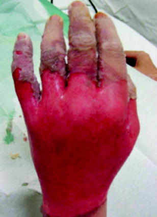
Superficial second degree burns usually present as blisters, appearing some hours after the accident. Once the blister is removed, the wound can be observed. Redness is uniform and pain is extreme, rarely allowing the physician to touch the lesion. Healing time is short, usually within the first 2 weeks, without aesthetic sequellae. The superficial dermis is exposed, without involving the basal membrane, which guarantees a quick healing in the superficial aspect of the skin (Figs. ).
Fig. 1.1
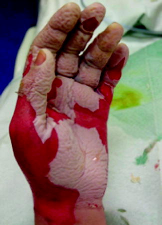
Early assessment of second degree burns over the dorsum of the hand. Blister has just been removed. Difficult to evaluate if deep. Reevaluate the next day and the day after
Fig. 1.2
Palmar aspect of the same hand. Same difficulty, but the fact that both aspects of the hand are involved is worse than when only one is involved
Fig. 1.3
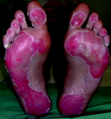
Sand burns of the palmar aspect of the feet after walking over a long distance on a hot beach. Second degree, superficial
Fig. 1.4
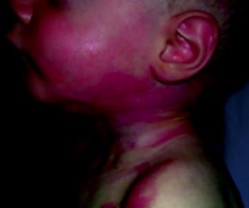
Fresh scald burns (second degree). Blister appearing progressively. Reevaluate after some hours before establishing a prognosis
Fig. 1.5
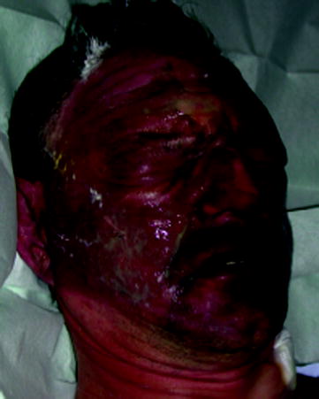
Fresh burns of the face. Ophtalmologic assessment. Removal of blisters is necessary before a proper assessment of the burns
Deep Second Degree
Deep second degree burns also present blisters, but after removal, the aspect is white or similar to patchwork. Sensibility to touch is not as important as in more superficial lesions, due to a partial destruction of sensory endings. Blanching of the skin under digital pressure cannot be obtained. These burns have a tendency to heal spontaneously, except in critical general conditions or if TBS burnt is extensive. The wound will stay unhealed or deteriorate and transform into a third degree burn. Usually, healing can be observed within 23 weeks, but as the deep dermis is exposed, a permanent scar will remain. These wounds can sometimes require an excision and a skin graft (Fig. ).
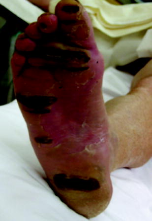
Fig. 1.6
Deep grill burns of the plantar aspect of the foot on a diabetic patient. Excision and grafting
Third Degree
Third degree burns are deep burns involving the subdermal structures. Extent in depth can be important, reaching aponeurosis or even bones. Lesions are sometimes circular on the limbs, a source of ischemia for the distal segments, necessitating emergency surgical procedures of discharge incisions to reestablish a normal distal blood flow. Lesions present with a white color and the tissues are hard. A black eschar will be observed after carbonization (Figs. ).

