• Complain

Ximena Wortsman (editor) - Dermatologic Ultrasound With Clinical and Histologic Correlations

Here you can read online Ximena Wortsman (editor) - Dermatologic Ultrasound With Clinical and Histologic Correlations full text of the book (entire story) in english for free. Download pdf and epub, get meaning, cover and reviews about this ebook. year: 2013, publisher: Springer Nature, genre: Home and family. Description of the work, (preface) as well as reviews are available. Best literature library LitArk.com created for fans of good reading and offers a wide selection of genres:

Romance novel Science fiction Adventure Detective Science History Home and family Prose Art Politics Computer Non-fiction Religion Business Children Humor

Choose a favorite category and find really read worthwhile books. Enjoy immersion in the world of imagination, feel the emotions of the characters or learn something new for yourself, make an fascinating discovery.

Ximena Wortsman (editor) Dermatologic Ultrasound With Clinical and Histologic Correlations

Dermatologic Ultrasound With Clinical and Histologic Correlations: summary, description and annotation

We offer to read an annotation, description, summary or preface (depends on what the author of the book "Dermatologic Ultrasound With Clinical and Histologic Correlations" wrote himself). If you haven't found the necessary information about the book — write in the comments, we will try to find it.

Significant technological advances have produced equipment that allows imaging of the skin with variable frequency ultrasound in previously unseen detail and provides a range of dynamic data that is currently unmatched by any other technology. Dermatologic Ultrasound with Clinical and Histologic Correlations is a comprehensive introduction to ultrasonography of the skin, nails, and scalp as it relates to the assessment and diagnosis of dermatologic diseases. It provides radiologists, sonographers, dermatologists, and physicians with interest in skin imaging with a concise understanding of the diagnosis of dermatologic conditions through extensive high-resolution gray scale and color Doppler ultrasound images and presents classical correlations of clinical dermatologic lesions with sonographic and histologic findings. Featuring more than 1700 images, this text-atlas provides an excellent starting point in learning about this topic.

Featuring contributions from world-renowned authorities in the field of superficial ultrasound imaging, the book reviews the technical considerations relating to color Doppler ultrasound of the skin; surveys the dermatologic entities that can be visualized with ultrasound imaging, such as cutaneous tumors, inflammatory diseases, hemangiomas and vascular malformations, melanoma, nail tumors, scalp diseases and cosmetic conditions; shows common simulators of cutaneous diseases; and discusses protocols for assessing common dermatologic conditions. Inclusion of clinical overviews, tips, and pitfalls enables a better understanding of the pathologies of the disorders and the methodological approach in assessing these entities.

Ximena Wortsman (editor): author's other books


Who wrote Dermatologic Ultrasound With Clinical and Histologic Correlations? Find out the surname, the name of the author of the book and a list of all author's works by series.


Dermatologic Ultrasound With Clinical and Histologic Correlations — read online for free the complete book (whole text) full work

Below is the text of the book, divided by pages. System saving the place of the last page read, allows you to conveniently read the book "Dermatologic Ultrasound With Clinical and Histologic Correlations" online for free, without having to search again every time where you left off. Put a bookmark, and you can go to the page where you finished reading at any time.

Light

Font size:

Reset

Interval:

Bookmark:

Make
Part 1
The Requisites
Ximena Wortsman (ed.) Dermatologic Ultrasound with Clinical and Histologic Correlations 2013 10.1007/978-1-4614-7184-4_1 Springer Science+Business Media New York 2013
1. Introduction to Color Doppler Ultrasound of the Skin
Diana Gaitini 1, 2
(1)
Unit of Ultrasound, Department of Medical Imaging, Rambam Health Care Campus, Haalya Hashnia 8, Haifa, 31096, Israel
(2)
Ruth and Bruce Rappaport Faculty of Medicine, Technion Israel Institute of Technology, Haifa, Israel
Diana Gaitini
Email:
1.1
1.2
1.3
Abstract
High-resolution gray scale and color Doppler ultrasound can be a useful tool in dermatology. Technical requirements and useful sonographic concepts are discussed in this chapter. An ultrasound glossary, tips and teaching points are provided to improve the understanding of this technique.
A comprehensive review of the basics of ultrasound focused on the requisites for skin, nail and scalp examinations
1.1 Introduction
Ultrasound has become a unique medical imaging tool in the investigation of dermatological diseases. By providing high-resolution gray scale images and blood flow information in real time, ultrasound can provide detailed anatomic and physiologic data of skin lesions and deeper soft-tissue changes. Lesion size in three dimensions-lengths, width and depth, morphology, inner structure -solid, cystic or mixed, homogeneous or inhomogeneous, foci of calcifications or necrosis, location, and extension can be diagnosed. By defining vascularity in real time, color and spectral Doppler ultrasound have been proven useful in the study of localized lesions of the skin. Interventional procedures such as tumor biopsy, collection drainage, foreign body removement, and needle localization of lesions can safely be performed under ultrasound guidance. The detailed anatomic information provided by sonography is useful in planning surgery. Magnetic resonance imaging (MRI) is frequently recommended for preoperative assessment although it requires the use of intravenous contrast media and can be less efficient in detecting tumors <3 mm in diameter. Ultrasound is a non-invasive and non-radiating test and as so, follow-up examinations can be performed without potential radiation damage. Finally, operator skills and knowledge of the clinical setting and the question being asked are important in ultrasound success.
1.2 Technical Considerations
Modern digital ultrasound systems utilize transducers with a broad range of frequencies (broad bandwidth) [). The operator should avoid pressing with the transducer because that may result in disappearing or false thinning of lesions.
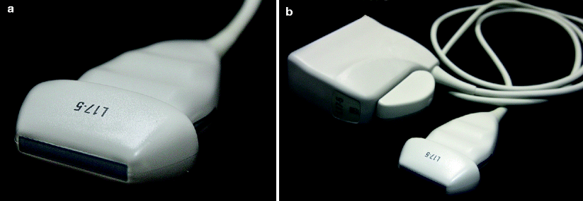
Fig 11 HVFUS linear array transducer a b High-resolution 50170 MHz - photo 1
Fig. 1.1
HVFUS linear array transducer. ( a , b ) High-resolution (5.017.0 MHz) transducers are used for superficial applications: small parts, breast, superficial vascular, and musculoskeletal including skin
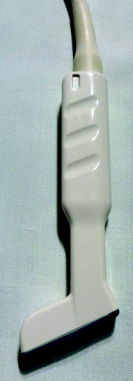
Fig 12 HVFUS linear array hockey stick transducer Versatile multi-frequency - photo 2
Fig. 1.2
HVFUS linear array hockey stick transducer. Versatile multi-frequency (up to 15 Mhz) compact hockey stick-shape transducer, with a high Doppler and color flow sensitivity. The hockey stick-shape allows complete contact with the skin surface, reducing scattering artifacts, making it a good choice for superficial and vascular imaging
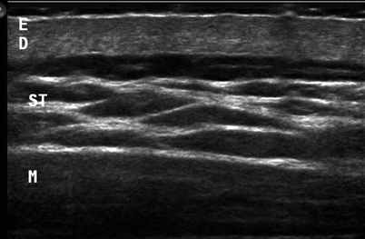
Fig 13 HVFUS image of skin layers and deeper structures Adjustments of focus - photo 3
Fig. 1.3
HVFUS image of skin layers and deeper structures. Adjustments of focus at a selected depth and selection of the transmission frequency provide a complete view of the skin and the deeper structures Frequencies in the upper range (1415 MHz) are used for skin layers and frequencies in the lower range (713 MHz) for the deeper tissues demonstration. E epidermis, D dermis, ST subcutaneous tissue, M muscle
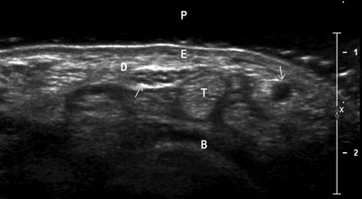
Fig 14 HVFUS image of superficial structures Axial scan at the level of the - photo 4
Fig. 1.4
HVFUS image of superficial structures. Axial scan at the level of the wrist. Variable frequency gives the best balance among spatial resolution and depth (2 cm penetration in this scan), generating good quality images of the skin ( E epidermis, D dermis), tendons ( T ), and bone ( B ). Subcutaneous venous and arterial blood vessels appear as echofree ducts ( upper arrow , ulnar artery). Nerves are seen as small hypoechoic dots surrounded by hyperechogenic tissue ( lower arrow , median nerve). A gel pad ( P ) was interposed between the transducer and the skin, to reduce near field artifacts
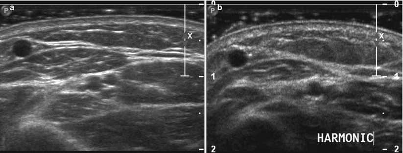
Modern equipment software includes capabilities for harmonic imaging, compound image, extended field of view (EFOV), and - D ultrasound . Tissue harmonic imaging reduces imaging artifacts caused by the interaction of the ultrasound beam with superficial structures or from aberrations at the edges of the beam profile. The artifact-producing signals are of low energy, insufficient to generate harmonic frequencies. Images generated exhibit reduced noise and improved spatial resolution (Fig. ). Three-dimensional (3D) imaging permits volume data to be displayed in multiple planes and allows accurate measurement of lesion volume.
Fig 15 ab Harmonic tissue imaging Split image at the same anatomic area - photo 5
Fig. 1.5 (a,b)
Harmonic tissue imaging. Split image at the same anatomic area ( a ) Compound scan. ( b ) Harmonic imaging. Harmonic imaging reduces near-field artifacts, caused by the interaction of the ultrasound beam with superficial structures. The generated images exhibit reduced noise and improved spatial resolution
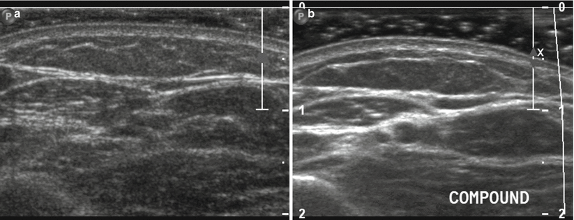
Fig 16 a b Spatial compounding imaging Split image at the same anatomic - photo 6
Fig. 1.6 (a, b)
Spatial compounding imaging. Split image at the same anatomic area ( a ) Fundamental scan. ( b ) Compounding imaging. Compound significantly improves contrast-to-noise ratio by reducing artifacts generated by the scattering of ultrasound from small tissue reflectors (speckle)
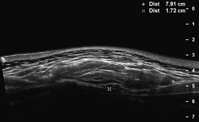
Fig 17 Extended field of view EFOV imaging EFOValso termed panoramic view - photo 7
Fig. 1.7
Extended field of view (EFOV) imaging. EFOValso termed panoramic view ( P in left image corner)allows acquisition and display of a panoramic image of large structures and the topographic anatomic relationships with the surrounding tissues, with no loss in resolution. In this case, an oval lesion in the subcutaneous tissue is seen, measuring 7.9 cm in length, compatible with a subcutaneous lipoma
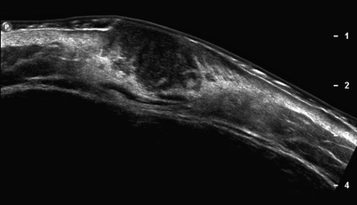
Fig 18 Panoramic EFOV view A malignant skin lesion dermatofibrosarcoma - photo 8
Fig. 1.8
Panoramic (EFOV) view. A malignant skin lesion (dermatofibrosarcoma) is presented. The tumor is hypoechogenic and poorly delimitated. Panoramic imaging shows lesion extension into the epidermis, dermis, and subcutaneous fat tissue
Next page
Light

Font size:

Reset

Interval:

Bookmark:

Make

Similar books «Dermatologic Ultrasound With Clinical and Histologic Correlations»

Look at similar books to Dermatologic Ultrasound With Clinical and Histologic Correlations. We have selected literature similar in name and meaning in the hope of providing readers with more options to find new, interesting, not yet read works.


Reviews about «Dermatologic Ultrasound With Clinical and Histologic Correlations»

Discussion, reviews of the book Dermatologic Ultrasound With Clinical and Histologic Correlations and just readers' own opinions. Leave your comments, write what you think about the work, its meaning or the main characters. Specify what exactly you liked and what you didn't like, and why you think so.