Usiakimi Igbaseimokumo - Brain CT Scans in Clinical Practice
Here you can read online Usiakimi Igbaseimokumo - Brain CT Scans in Clinical Practice full text of the book (entire story) in english for free. Download pdf and epub, get meaning, cover and reviews about this ebook. year: 2019, publisher: Springer, genre: Home and family. Description of the work, (preface) as well as reviews are available. Best literature library LitArk.com created for fans of good reading and offers a wide selection of genres:
Romance novel
Science fiction
Adventure
Detective
Science
History
Home and family
Prose
Art
Politics
Computer
Non-fiction
Religion
Business
Children
Humor
Choose a favorite category and find really read worthwhile books. Enjoy immersion in the world of imagination, feel the emotions of the characters or learn something new for yourself, make an fascinating discovery.

- Book:Brain CT Scans in Clinical Practice
- Author:
- Publisher:Springer
- Genre:
- Year:2019
- Rating:3 / 5
- Favourites:Add to favourites
- Your mark:
Brain CT Scans in Clinical Practice: summary, description and annotation
We offer to read an annotation, description, summary or preface (depends on what the author of the book "Brain CT Scans in Clinical Practice" wrote himself). If you haven't found the necessary information about the book — write in the comments, we will try to find it.
Usiakimi Igbaseimokumo: author's other books
Who wrote Brain CT Scans in Clinical Practice? Find out the surname, the name of the author of the book and a list of all author's works by series.


Brain CT Scans in Clinical Practice — read online for free the complete book (whole text) full work
Below is the text of the book, divided by pages. System saving the place of the last page read, allows you to conveniently read the book "Brain CT Scans in Clinical Practice" online for free, without having to search again every time where you left off. Put a bookmark, and you can go to the page where you finished reading at any time.
Font size:
Interval:
Bookmark:
Taking a practical approach to clinical medicine, this series of smaller reference books is designed for the trainee physician, primary care physician, nurse practitioner and other general medical professionals to understand each topic covered. The coverage is comprehensive but concise and is designed to act as a primary reference tool for subjects across the field of medicine.
More information about this series at http://www.springer.com/series/13483

This Springer imprint is published by the registered company Springer Nature Switzerland AG.
The registered company address is: Gewerbestrasse 11, 6330 Cham, Switzerland
Interpretation of the emergency CT brain scan is a visual art. Comparison is made between the image in front of you and a reference image. For the experienced person this reference image is imprinted in the mind; therefore comparison is quick. For the beginner you can either carry several examples of every possible appearance of normal and abnormal scans to compare with or read this book! This book contains a few proven ways of quickly learning to interpret a brain CT scan, irrespective of your previous experience.
The radiologists experience is related to the number of hours he or she has spent looking at CT scans. The radiologist conveys his evaluation of the CT scan in words that often come in a particular sequence and combination. This book is about helping you to rapidly understand and confidently use the same language used by the radiologist.
The difference is that whereas the radiologist aims for perfection, you aim for functionality. For instance, it will be acceptable and clinically safe if an intern physician looks at the brain CT scan in Fig. and can make a judgement of the urgent action required like ABCs ( A irway, B reathing and C irculation with c- s pine) and call a neurosurgeonimmediately. This is lifesaving and efficient without the need for a long list of differential diagnoses before deciding on this action. Since the first edition, image presentation and storage has moved from films to digital format. The author has seamlessly introduced this new platform in explaining the concepts of interpreting the emergency brain CT scan. Therefore, the skill to act decisively about the CT scan in front of you can be acquired in a very short time. And the author has reduced that time to a few hours using this book!
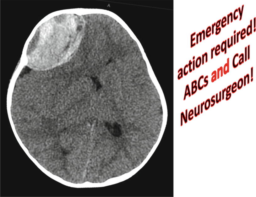
Epidural hematoma! Emergency action required! ABCs and call neurosurgeon!
An illustrated guide for ER physicians, trauma surgeons, primary care physicians, residents, medical students, nurses and other care givers
Across emergency rooms all over the world, thousands of patients are referred for brain CT scans daily. A radiologist often has to interpret the scan or a consultation has to be made to a neurosurgeon to review the scan. Most of this happens late at night and is a significant source of discontent. Thus, having frontline physicians to be proficient in interpreting the emergency brain CT scan improves the efficiency of the whole pathway of care and is potentially lifesaving as time is of the essence for many patients with severe brain injury or stroke.
Underlying all of the above and the primary reason for writing this book is that the skill required to determine an immediate life-threatening abnormality in a brain CT scan is so basic and can be learned in a short time by people of various backgrounds and certainly by all physicians. Indeed, the emergency head CT scan is comparable to an electrocardiogram in usefulness and most definitely as easy to learn. This book is therefore written for caregivers the world over to demystify the emergency CT brain scan and to empower them to serve their patients better. It is obvious to me from the responses from people I have had opportunity to teach this subject that not only is there a desire to learn this basic skill but also people learn it quickly and wonder why it has not been presented so simply before.
It is to fulfil this need and to reach a wider number that I have put together these basic, proven steps in the interpretation of emergency brain CT scan for ER physicians, trauma surgeons, primary care physicians, nurses, medical students and other primary caregivers.
Since the first edition, the transition from X-ray film-based delivery of images to electronic delivery (e.g. PACS) has become more widespread. However, the examination of individual slices to reconstruct a three-dimensional image remains the cornerstone of CT imaging; hence this volume has retained the present format while recognizing the ability of the non-radiologist provider to manipulate individual images including enlarging and changing the window level. This technology is by no means universal and therefore not assumed in the description of images in this text.
My heartiest gratitude goes to my wife Ebitimi and my kids Gesiye, Ilayefa and Binaere who volunteered the real cost in time to prepare this book. My eternal gratitude to the IsounsTurner and Miriamfor spiritual, financial and intellectual support on this and every other project I ever embarked upon, thank you.
Font size:
Interval:
Bookmark:
Similar books «Brain CT Scans in Clinical Practice»
Look at similar books to Brain CT Scans in Clinical Practice. We have selected literature similar in name and meaning in the hope of providing readers with more options to find new, interesting, not yet read works.
Discussion, reviews of the book Brain CT Scans in Clinical Practice and just readers' own opinions. Leave your comments, write what you think about the work, its meaning or the main characters. Specify what exactly you liked and what you didn't like, and why you think so.