Gerbail T. Krishnamurthy and S. Krishnamurthy Nuclear Hepatology A Textbook of Hepatobiliary Diseases 10.1007/978-3-642-00648-7_1 Springer-Verlag Berlin Heidelberg 2009
1. Morphology and Microstructure of the Hepatobiliary System
Gerbail T. Krishnamurthy 1 and Shakuntala Krishnamurthy 1
(1)
Tuality Community Hospital, 97123 Hillsboro, OR, USA
Abstract
Liver and gallbladder disease is a common entity around the world and according to the World Health Organization estimation accounts for 46.1% of global disease [1]. Liver transplantation for end-stage liver disease, segmental resection for tumor, and therapeutic interventional maneuvers has made it necessary to have thorough knowledge of the morphology of the liver and biliary system in much greater detail than ever before [2]. With the widespread application of segmental liver resection for living donor liver transplantation or radioablation of liver tumors, thorough knowledge of internal structures is critical for radiologists, nuclear medicine physicians, and surgeons. Since the publication of the first edition of our book in 2000, many advances in liver disease therapy have taken place, making it necessary to provide more detailed anatomic and histopathological information. Anatomical details of the vascular and ductal structures are well depicted on a multi-detector computer tomography (MDCT), magnetic resonance cholangiopancreatography (MRCP), and endoscopic retrograde cholangiopancreatography (ERCP), enabling identification of the third or fourth order branches on the images. Such detailed structural information is necessary for surgeons for segmental resection of the liver.
1.1 Morphology
Liver and gallbladder disease is a common entity around the world and according to the World Health Organization estimation accounts for 46.1% of global disease []. With the widespread application of segmental liver resection for living donor liver transplantation or radioablation of liver tumors, thorough knowledge of internal structures is critical for radiologists, nuclear medicine physicians, and surgeons. Since the publication of the first edition of our book in 2000, many advances in liver disease therapy have taken place, making it necessary to provide more detailed anatomic and histopathological information. Anatomical details of the vascular and ductal structures are well depicted on a multi-detector computer tomography (MDCT), magnetic resonance cholangiopancreatography (MRCP), and endoscopic retrograde cholangiopancreatography (ERCP), enabling identification of the third or fourth order branches on the images. Such detailed structural information is necessary for surgeons for segmental resection of the liver.
1.1.1 Embryology
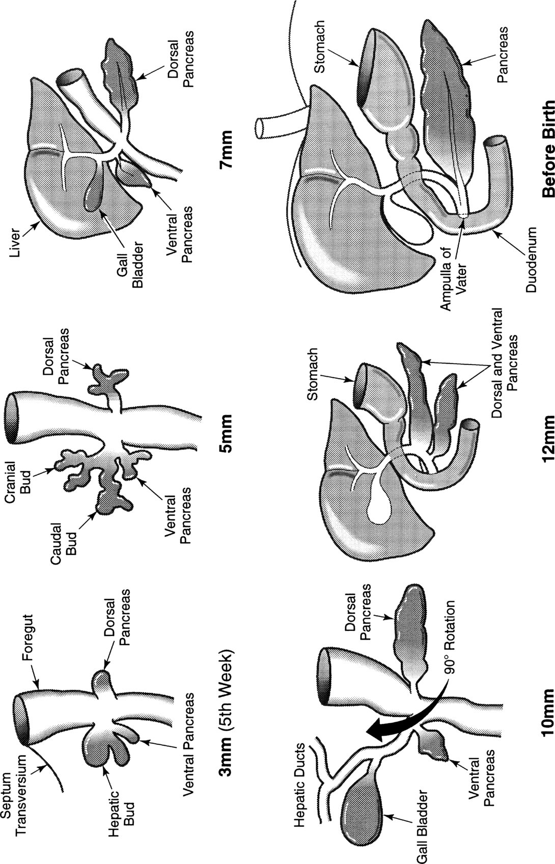
The liver and biliary systems develop from an endodermal bud that arises during the 5th week of intrauterine life when the embryo is about 3 mm in length []. This rotation brings the common bile duct posterior to the duodenum. Many congenital abnormalities around this region are secondary to mal-rotation at this junction. The caudate lobe arises separately close to the inferior vena cava, independent of the right and left hepatic lobes. The liver begins bile secretion by the 12th intrauterine week, thus completing the formation of the hepatobiliary system, the most complex metabolic factory in the human body. In adults, the liver weighs about 1,5001,800 g and forms about 1/50 of the body weight. In children, however, it forms a relatively much larger fraction (1/20) of the total body weight.
Fig. 1.1.1
Embryology of the hepatobiliary system. The hepatic bud arises from the endoderm of the primitive foregut at its junction with the midgut when the embryo is about 3 mm in length. The hepatic bud divides into cranial (pars hepatica) and caudal (pars cystica) branches when the embryo reaches 5 mm size. The ventral pancreas arises from the pars cystica, which later gives rise to the biliary system. After a 90 clockwise rotation (10 mm embryo), the ventral pancreas fuses with the dorsal pancreas. This rotation brings the common bile duct posterior to the duodenum (12 mm). The common bile duct opens into the duodenum at the postero-medial wall at an elevation called the papilla
1.1.2 Liver Lobes and Surfaces
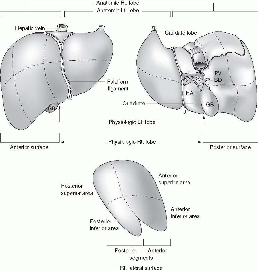
Most of the liver is situated in the right upper quadrant of the abdomen underneath the right hemi-diaphragm, with the superior border situated at the level of the right fifth intercostal space. Liver consists of five surfaces: anterior, posterior, right lateral, superior, and inferior. The anterior, right lateral, and posterior surfaces are smooth, and superior and inferior surfaces are rough with grooves and fissures for entry and exit of the vascular and biliary structures (Fig. ].
Fig. 1.1.2
Surfaces, segments, and lobes of the liver. The anatomic left lobe (divided by the falciform ligament) is much smaller than the anatomic right lobe, but the physiologic left lobe is much larger than the anatomic left lobe. The quadrate lobe, which forms a part of the anatomic right lobe, forms a part of the physiologic left lobe. Liver consists of three lobes (including the caudate lobe), four segments, and eight areas. HA hepatic artery, BD bile duct, PV portal vein, GB gallbladder
The falciform ligament, the marker of the anatomic right and left lobes, lies to the left of the deep fissure (Srg-Cantlie line) that divides the liver into physiologic right and left lobes. The caudate lobe situated posteriorly and quadrate lobe situated anteriorly form part of the anatomic right lobe, but both of these structures belong to the physiologic left lobe (Fig. ). The anatomic right lobe is approximately six times larger (85%) than the anatomic left lobe (15%), whereas the physiologic right lobe is 70% and physiologic left lobe 30% in size. Thus, the physiologic left lobe is much larger than its anatomic counterpart. It is important, therefore, to be very specific while describing lobes of the liver, whether one is referring to the anatomic or to the physiologic division. Throughout this book, we will refer to the physiologic division unless otherwise mentioned.
1.1.3 Segments and Areas
The liver is divided into lobes, segments, and areas. The division is made on the basis of either the vascular [].
Table 1.1.1
Nomenclature for hepatic segments and lobes as adopted by different authors
Liver lobes | Hjortsjo (5) | Healey and Schroy (8) | Couinaud (6) | Bismuth (9) |
Caudate lobe | Dorsal segment | Lobus caudatus | I | I |
Left lobe | Dorso-lateral segment Ventro-lateral segment Central segment Dorso-ventral segment | Lateral superior area Lateral inferior area Medial superior area Medial inferior area | II III IV IV | II III IVA IVB |
Right lobe | Ventro-caudal segment Dorso-caudal + intermedio-caudalDorso-cranial + intermedio-cranialVentro-cranial | Anterior-inferior area Posterior-inferior area Posterior-superior area Anterior-superior area |
