Springer Nature Singapore Pte Ltd. 2018
Hongda Wang and Guohui Li (eds.) Membrane Biophysics
1. Composition and Function of Cell Membranes
Mingjun Cai 1, Jing Gao 1 and Hongda Wang 1
(1)
State Key Laboratory of Electroanalytical Chemistry, Changchun Institute of Applied Chemistry, Chinese Academy of Sciences, No. 5625, Renmin Street, Changchun, 130022, Jilin, Peoples Republic of China
1.1 Introduction
The cell membrane encloses the cell, defines its boundaries, and maintains the essential differences between the cytosol and the extracellular environment. Inside eukaryotic cells, the membranes of the nucleus, endoplasmic reticulum, Golgi apparatus, mitochondria, and other membrane-enclosed organelles maintain the characteristic differences between the contents of each organelle and the cytosol. All these membranes form enclosed structure and a related net called cytoplasmic membrane system. Ion gradients across membranes, established by the activities of specialized membrane proteins, can be used to synthesize adenosine triphosphate (ATP), to drive the transport of selected solutes across the membrane, or as in nerve and muscle cells, to produce and transmit electrical signals. In all cells, the cell membrane also contains proteins those act as sensors of external signals, allowing the cell to change its behavior in response to environmental cues, including signals from other cells; these protein sensors, or receptors, transfer information rather than molecules across the membrane. Cell membranes are dynamic, fluid structures, and most of their molecules move about in the plane of the membrane.
1.2 Composition of Cell Membranes
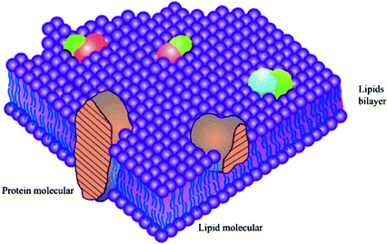
Despite their different functions, all biological membranes have a general structure: a very thin film of lipid and protein molecules, linking together mainly by non-covalent interactions [].
Fig. 1.1
Fluid-mosaic membrane model of cell membrane structure. Reprinted from Ref. [], Copyright 2014, with permission from Elsevier
1.2.1 Lipids
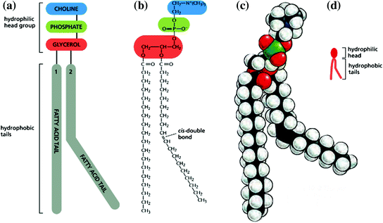
Lipid bilayer constitutes the fundamental structure of all the cell membranes. The lipids in cell membranes combine two very different properties in a single molecule: Each lipid has a hydrophilic (water-loving) head and a hydrophobic (water-fearing) tail. The most abundant lipids in cell membranes are the phospholipids, which have a phosphate-containing hydrophilic head linked to a pair of hydrophobic tails (Fig. ). The bilayer structure is maintained by hydrophobic and Van der Waals force.
Fig. 1.2
Structure of phosphatidylcholine, a typical phospholipid molecule. a Scheme of molecular structure, b Formula, c Space-filling model, and d Symbol. Reprinted from Ref. [] by permission of Garland Science Taylor & Francis Group
Lipid molecules constitute about 50% of the mass of most animal cell membranes, and nearly all of the remainder are proteins. There are approximately 5 106 lipid molecules in a 1 1 m area of lipid bilayer or about 109 lipid molecules in the cell membrane of a small animal cell. Lipids contained in typical cell membrane are phospholipids (include phosphoglycerides, sphingolipids) and steroids.
1.2.1.1 Phosphoglycerides
Phosphoglycerides derivatives of glycerol 3-phosphate are the most abundant class of lipids in most membrane. A typical phosphoglyceride molecule consists of a polar head attached to the phosphate group, and a hydrophobic tail composed of two fatty acyl chain esterified to the two hydroxyl groups in glycerol phosphate. The fatty acyl chains may differ in the number of carbons (they normally contain between 14 and 24 carbon atoms) and their degree of saturation (0, 1, or 2 double bonds). The length and the saturation degree of the fatty acid tails are important in regulating the fluidity of the membrane (Fig. ).
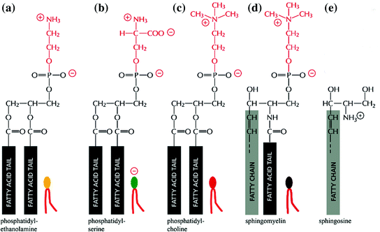
Phosphoglycerides are classified according to the nature of their head groups. In phosphatidylcholines, for example, the head group consists of choline; a positively charged alcohol is esterified to the negatively charged phosphate. In other phosphoglycerides, an OH-containing molecule such as ethanolamine, serine, or the sugar derivative inositol is linked to the phosphate group. The negatively charged phosphate group and the positively charged or the hydroxyl groups on the head group interact with water (Fig. ).
Fig. 1.3
Four major phospholipids in mammalian cell membranes. Different head groups are represented by different colors in the symbols. The lipid molecules shown in ( a c ) are phosphoglycerides, which are derived from glycerol. The molecule in ( d ) is sphingomyelin, which is derived from sphingosine ( e ) and is therefore a sphingolipid. Note that only phosphatidylserine carries a net negative charge, and the other three are electrically neutral at physiological pH, carrying one positive and one negative charge. Reprinted from Ref. [] by permission of Garland Science Taylor & Francis Group
1.2.1.2 Sphingolipids
The second class of membrane lipids is sphingolipid. Sphingolipid is derived from sphingosine rather than glycerol (Fig. ).
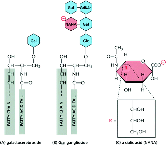
Fig. 1.4
Structure of glycolipid molecules in cell membrane. a Galactocerebroside is called a neutral glycolipid because the sugar that forms its head group is uncharged. b A ganglioside always contains one or more negatively charged sialic acid moiety. There are various types of sialic acid; in human cells, it is mostly n -acetylneuraminic acid, or NANA), whose structure is shown in ( c ). Whereas in bacteria and plants almost all glycolipids are derived from glycerol, as are most phospholipids, in animal cells almost all glycolipids are based on sphingosine, as is the case for sphingomyelin (see Fig. ] by permission of Garland Science Taylor & Francis Group
Table 1.1
Compares the lipid compositions of several biological membranes
Lipid | Percentage of total lipid by weight |
Liver cell membrane | Red blood cell membrane | Myelin | Mitochondrion (inner and outer membranes) | Endoplasmic reticulum | E. colibacterium |
Cholesterol | | | | | | |
Phosphatidylethanolamine | | | | | | |
Phosphatidylserine | | | | | | Trace |
Phosphatidylcholine | | | | | | |
Sphingomyelin | | | | | | |
Glycolipids | | | | Trace | Trace | |

