1.1 Introduction
To answer many questions about how a fungal cell functions, it is necessary to examine cellular and subcellular events such as the behaviors and locations of specific organelles and proteins. For this purpose, there are a variety of fluorescence -imaging modalities that are commonly used, such as wide-field microscopy, laser-scanning confocal microscopy (LSCM) (Czymmek et al. ). Though a working microscope was produced in 1955, the technological advances required to construct a practical research instrument delayed bringing it to market until the mid-1980s. Confocal microscopy has since become a widely used technique for imaging an extensive range of fluorescently labeled biological specimens, including fungi. Over the years, major improvements have been made in the technology applied in recording and displaying confocal images with high resolution and in determining the availability of more photo-stable fluorophores. Initially, confocal microscopy applications were used mainly in fixing images of fluorescently labeled specimens using, for example, immunofluorescence protocols. The imaging of living organisms has become more applicable to a wider range of specimens with the introduction of fluorescent proteins , use of fluorescent vital dyes, and improved confocal systems (e.g., optics, light path, scanning strategies, and detector electronics) that have reduced photobleaching and phototoxicity. Live-cell imaging techniques provide a means for documenting organelle and macromolecular dynamics with increased spatial and temporal resolutions. Other advantages of confocal microscopy are the experimental approaches for multiple fluorescent labels and multidimensional microscopy.
The optical section is the basic image data obtained from a confocal microscope and refers to its ability to produce images along the z-axis (through the sample) of fluorescently-labeled specimens without resorting to physical sectioning of the sample (Fig. ). Optical sections are produced in LSCM by scanning a region of interest of the specimen point-by-point with a focused laser beam, and using a pinhole aperture in front of the detector to remove out-of-focus fluorescent signal from above and below the focal plane of interest. Optical paths of confocal microscopy are designed in such a way that the laser beam is focused within the specimen and the resultant fluorescent signal is refocused at a pinhole in front of the detector (thus, the two focal points, making it confocal).The power of this approach lies in its ability to clearly image structures at discrete Z levels within an intact biological specimen.
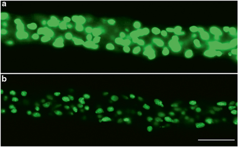
Fig. 1.1
Comparison of ( a ) a wide-field epifluorescence and ( b ) a confocal microscopy image of nuclei labeled with histone H1 fused to GFP in the filamentous fungus Neurospora crassa . Scale Bar=10 m. (Images by Olga A. Callejas-Negrete)
1.2 The Confocal Microscopy System
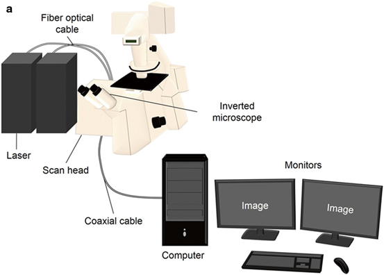
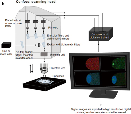
The confocal microscopy system includes integrated optical and electronic devices such as a research grade microscope , multiple sources of coherent light (laser beams), a confocal scan head, a computer with one or two monitors and software for image acquisition, processing and analysis (Fig. ).
Fig. 1.2
Typical components of a laser-scanning confocal microscopy system. a Includes a research-grade microscope, upright or inverted, a scan head, laser beams, computer, and monitors. b Diagram of the confocal scanning head. It shows the laser input, the excitation filters and the emission filters, galvano mirrors and detectors. (Artwork by Arianne Ramirez-del Villar)
Confocal microscopy was developed to remove out-of-focus light (haze) from the objective focal plane. How does it work?
First, the specimen must be illuminated with a specific wavelength of light to excite the fluorophore. Laser beams are used to illuminate the specimen through the objective lens, which is also a receptacle for the specimen-emitted light; thus, this lens functions as both a condenser and an objective. Dichroic mirrors, prisms, diffraction gratings, or tunable filters are used to obtain specific spectral excitation and/or emission signals. In LSCM, the laser beam fills the objective aperture and forms an intense diffraction-limited spot that is raster-scanned quickly from side to side and from top to bottom pixel by pixel across the entire specimen in a process called point-scanning.
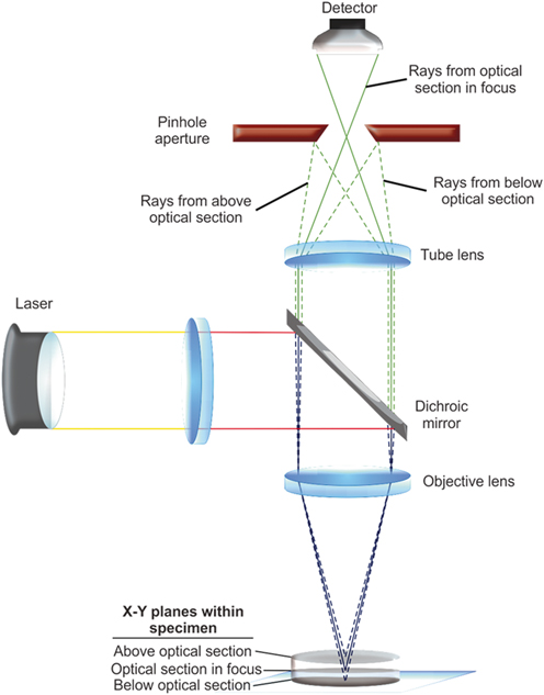
As previously mentioned, the light emitted from the specimen is collected by the objective lens and then follows the microscopes optical path, reaching the pinhole aperture in front of the detector required to produce confocal images. The pinhole aperture is placed at a precise distance from the detector where the image-forming train of light is focused (Fig. ).
Fig. 1.3
Diagram of the light path in a confocal microscopic system. Pinhole aperture is in front of the detector in order to eliminate out-of-focus light. The image recorded therefore comes from one thin optical plane within the specimen ( solid line ) and the light above and below is blocked ( discontinuous line ). (Artwork by Fausto M. Villavicencio-Aguilar)
There are also alternative ways of collecting emitted light from the specimens when we are interested in a specific wavelength range (user definable). Emitted light can be separated into spectral bands using the method called spectral imaging, achieved by utilizing prisms, diffraction gratings, or tunable filters to obtain a specific spectral emission signal (Haraguchi et al. ).
Excitation and emission light paths are separated by a dichroic mirror or acoustooptic beam splitter before the light passes through the pinhole aperture to reach the scan head. The filtered light arrives at a galvano-scanning mirror that is constantly oscillating, and sends the signal to the detecting PMT or a more photon-sensitive GaAsP detector at an image plane in a wide-field fluorescence microscope (light path: filters-pinhole-Galvano mirror-detection device). The detectors do not perceive an image per se, but produce a voltage that corresponds to the intensity of incident light (photons) and the software digitizes the signal and displays it as an image on the monitor (Fig. ).