Bedside Ultrasound Level 1
ISBN: 978-0-9919566-8-5
ISBN: 978-1-9992477-2-0 (e-book)
Copyright
Text 2013 Peter Steinmetz
Illustrations 2013 Sharon Oleskevich
ALL RIGHTS RESERVED. This book is protected by domestic and international copyright laws, making it illegal for anyone to photocopy, scan, reprint, email, give away, sell or otherwise distribute copies, whether in digital or physical format. No part of this document may be reproduced or transmitted in any form without the prior written consent of the publisher. Copyright infringement is a criminal offense with fines up to $5,000 for all individual infringement and $20,000 for each commercial infringement.
First Edition
first printing, June 2013
second printing, October 2013
third printing, February 2014; revisions include new figures (), and correction of minor typographical errors.
Second Edition
first printing, August 2018; this second edition includes improved and updated chapters, and the Canadian Point of Care Ultrasound Society guidelines. We also added false-positives and false-negatives for each of the indications. Overall, this book includes new text (44 pages), new illustrations to better illustrate important concepts (29 figures), and new instructional videos (2 videos).
The information in this book is designed to provide helpful information on the subjects discussed. While best efforts have been made to provide accurate information that is in accord with current standards of practice, the author, editor, or publisher make no warranty with respect to the accuracy or completeness of the contents. Application of the information in a particular situation remains the professional responsibility of the physician. Any likeness to actual persons in the case studies is strictly coincidental. References are provided for informational purposes only and do not constitute endorsement of websites or other sources.
Scientific editor
Sharon Oleskevich PhD
Web manager for online videos
John D. Clements PhD
Proofreader
Christiane de Brentani BA
Printer
IngramSpark / Lightning Source Inc.
Publisher
A-line Press, Montreal, Canada
P REFACE
This introductory handbook is a practical reference for healthcare workers starting to apply bedside ultrasound in their daily practice. It aims to articulate the practical and cognitive skills necessary to effectively use this tool.
In this second edition, we have improved and updated each chapter, and added the Canadian Point of Care Ultrasound Society guidelines. We also added false-positives and false-negatives for each of the indications. Overall, this book includes new text (44 pages), new illustrations to better illustrate important concepts (29 figures), and new instructional videos (2 videos).
The instructional ultrasound videos can be accessed at:
bedsideultrasoundlevel1.com
D EDICATION
"My only question is, why haven't we done this sooner?"
Arnold Steinberg, 2012
This textbook is dedicated to the late Arnold Steinberg whose vision and support were instrumental in establishing the bedside ultrasound curriculum at McGill University.
A BOUT THE AUTHOR
Dr. Peter Steinmetz is an Assistant Professor in the Department of Family Medicine at McGill University. He founded and leads undergraduate bedside ultrasound teaching at McGill University. As an executive board member of the Canadian Point of Care Ultrasound Society, he helps set national certification standards for ultrasound use in the family physicians office. He has published on the accuracy and skill acquisition of medical students using bedside ultrasound. He has also been invited to teach bedside ultrasound in Asia (Thailand) and Africa (Rwanda) and co-directed the World Congress of Ultrasound in Medical Education.
Dr. Steinmetz currently works at a community health clinic where point of care ultrasound is actively used in a family medicine setting.
A CKNOWLEDGEMENTS
There are countless people who deserve mention and a word of thanks for inspiring this second edition. They include the hundreds of McGill medical students who used this book to learn ultrasound and gave constructive feedback over the last five years of our groundbreaking bedside ultrasound curriculum. I am also thankful to the Canadian Point of Care Ultrasound Society for providing clear guidelines for bedside ultrasound use by Canadian clinicians as outlined at the end of relevant chapters. For the French edition, I am grateful to Dr. Claude Topping for his precise translations as well as important additions to the text itself.
A special thanks to Sharon Oleskevich for countless hours of editing, design, organization, writing, and general good advice.
P.S.
C ONTENTS
ULTRASOUND BASICS
1.1What is ultrasound?
Ultrasound is a sound wave that oscillates with a frequency greater than 20,000 Hz (20,000 cycles per second or 20 kHz). The human ear can detect sound waves with frequencies between 0.02-20 kHz. Sound waves above 20 kHz, such as those generated by dog whistles, bat calls, and ultrasound machines, cannot be interpreted as sound by the human ear. These high frequency sound waves are called ultrasound.
It is useful to understand some basic concepts about ultrasound in order to correctly interpret the images generated by the bedside ultrasound machine.
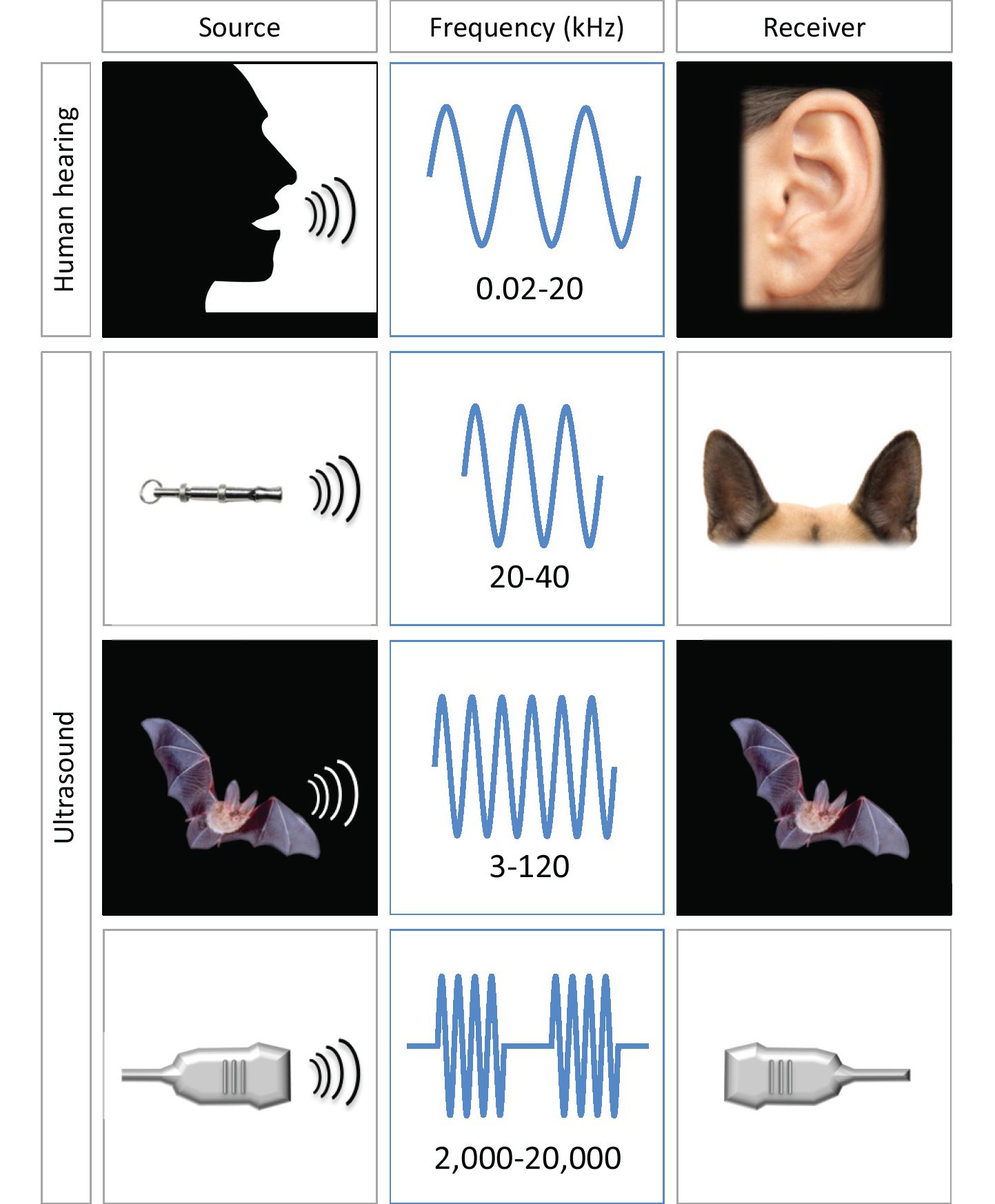
Figure 1.1 Different sound wave frequencies.
The human ear can interpret sound waves with frequencies up to 20 kHz. Sound waves with frequencies above 20 kHz are termed ultrasound. Ultrasound probes emit frequencies from 2,000-20,000 kHz (2-20 MHz).
1.2How do ultrasound probes send and receive ultrasound?
Ultrasound probes have two functions: to send and receive ultrasound. An ultrasound probe spends 1% of its time sending sound waves and 99% of its time receiving sound waves.
The probe starts by sending out bursts of ultrasound. When ultrasound encounters a structure, it reflects off that structure and returns to the probe. The reflected ultrasound is interpreted and expressed as an image on the ultrasound machine monitor.
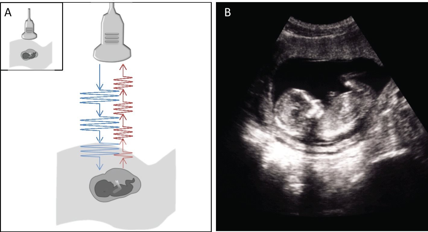
Figure 1.2 Ultrasound probes send and receive ultrasound.
A. Schematic demonstrating a probe sending sound waves (blue) and receiving the reflected returning sound waves (red).
B. The returning sound waves are detected by the probe and translated into an ultrasound image.
1.3How does ultrasound behave travelling through tissue?
Attenuation
Attenuation describes the dampening of the amplitude of ultrasound as it travels through tissue. Attenuation occurs as ultrasound energy is converted into heat, absorbed by a structure, reflected back to the probe, or scattered away from the probe.

