Page List

Published in 2019 by The Rosen Publishing Group, Inc.
29 East 21st Street, New York, NY 10010
Copyright 2019 by The Rosen Publishing Group, Inc.
First Edition
All rights reserved. No part of this book may be reproduced in any form without permission in writing from the publisher, except by a reviewer.
Library of Congress Cataloging-in-Publication Data
Names: Brezina, Corona, author.
Title: Jump-starting a career in ultrasound and sonography / Corona Brezina.
Description: First edition. | New York : Rosen YA, 2019. | Series: Health care careers in 2 years | Audience: Grades 712. | Includes bibliographical references and index.
Identifiers: LCCN 2018011693| ISBN 9781508185116 (library bound) | ISBN 9781508185109 (pbk.)
Subjects: LCSH: Ultrasonic imagingVocational guidanceJuvenile literature. | Diagnostic ultrasonic imagingVocational guidanceJuvenile literature.
Classification: LCC RC78.7.U4 B74 2019 | DDC
616.07/543023dc23
LC record available at https://lccn.loc.gov/2018011693
Manufactured in the United States of America
CONTENTS
F or an expectant mother, the day of the midpregnancy ultrasound procedure brings feelings of excitement and anxiety. It represents a progress report on the pregnancy and the health of the developing fetus.
The ultrasound is performed by a diagnostic medical sonographer, who scans the mothers belly while observing the image on a monitor. To most observers, the screen appears to be an abstract jumble of different shades of gray. To the sonographer, however, the image conveys crucial medical information that she can decipher at a glance. (The field of sonography is largely dominated by women, and most sonographers who work in womens health specialties are women, according to labor force statistics compiled by the US Bureau of Labor Statistics [BLS] in January 2018.) She may point out the position and movement of the fetus on the screen to the mother. Shell monitor the babys heart and determine whether its a boy or a girl, if the position of the fetus will allow and the mother wishes to be told.
Many mothers remember this ultrasound procedure as their first baby picture. But for the sonographer, the most important aspects concern the health of the mother and fetus. The sonographer checks for abnormalities in the babys anatomy and performs measurements that will confirm the babys growth and health. She will also confirm the health of the mothers reproductive organs.
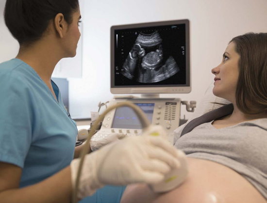
An ultrasound of a fetus can confirm that the pregnancy is proceeding normally. Sometimes, however, it reveals abnormalities that may affect the health of the unborn baby.
Most mothers go home after the ultrasound thrilled to share the news that the baby is beautiful and healthy. In some cases, however, the sonographer may observe an abnormality that requires further scans or tests. If a serious condition is diagnosed, it may require treatment during pregnancy or intensive monitoring.
Although fetal ultrasounds are the best-known application of sonography, diagnostic medical sonographers perform many different types of ultrasound procedures on various areas of the body. Sonographers analyze abnormalities seen in ultrasound images to diagnose medical conditions. They often say that they have to act as detectives during the course of their work to figure out the correct diagnosis.
Sonography is a rewarding health care career that pays well after the completion of a two-year degree. Health care is an expanding sector of the economy, and sonography in particular is expected to experience job growth. The field of sonography is not for everyoneit requires a combination of people skills, technical ability, critical thinking, attention to detail, and mental and physical stamina. But many diagnostic medical sonographers consider it an exciting and fulfilling profession in which their work can play a pivotal role in a patients diagnosis, treatment, and recovery.
D iagnostic medical sonographers, also called ultrasonographers, ultrasound technicians, or ultrasound technologists, operate medical equipment that obtain images of a patients body that are used for diagnosis. Sonography, sometimes called ultrasonography, is used to detect a broad range of different medical conditions.
Ultrasound has been utilized for various purposes for decades. Medical ultrasound imaging technology is based on the same principles that are applied in SONAR (SOund Navigation And Ranging) instruments. SONAR was employed by warships during World War II (1939 1945) to detect enemy submarines. After the war ended, scientists began investigating ways that ultrasound could be used to scan the human body. The first modern medical ultrasound equipment was introduced in the 1980s, taking advantage of advances in computing. Today, ultrasound is used in a variety of applications other than medicine, from motion sensors to industrial processing. A form of it even occurs in the natural worldbats avoid obstacles using echolocation, in which they bounce sound waves off of solid objects. Some marine mammals such as dolphins, porpoises, and toothed whales use echolocation, too.
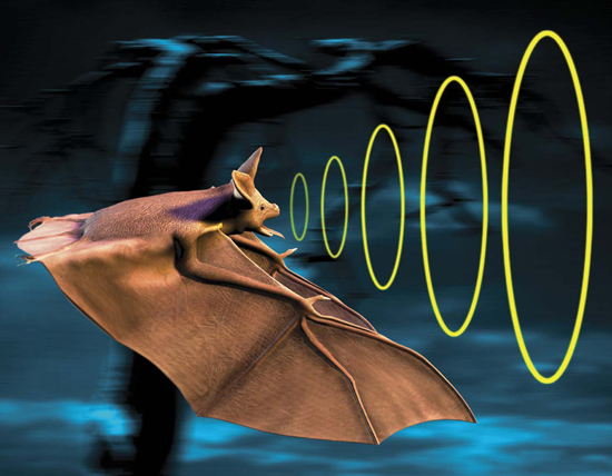
Bats navigate and locate prey by echolocation, emitting high-pitched sounds that bounce off solid objects and are detected by their large, sensitive ears.
What Is Sonography?
Sonographers use instruments that direct high-frequency sound waves into the body. The sound waves that are used for ultrasound procedures are too high in pitch for humans to detect them.
The most commonly used medical application of ultra-sound is diagnostic ultrasound, which produces images of the body. The sound waves used in diagnostic ultrasound are produced by a probe called a transducer, which is connected to the ultrasound machine. Before beginning the procedure, the sonographer coats the area of the body being scanned with ultrasound gel, which ensures close contact between the transducer and the skin. (In some cases, the probe will instead be placed inside the body.) The sonographer rests the transducer on the skin and views the resulting image on a monitor throughout the procedure.
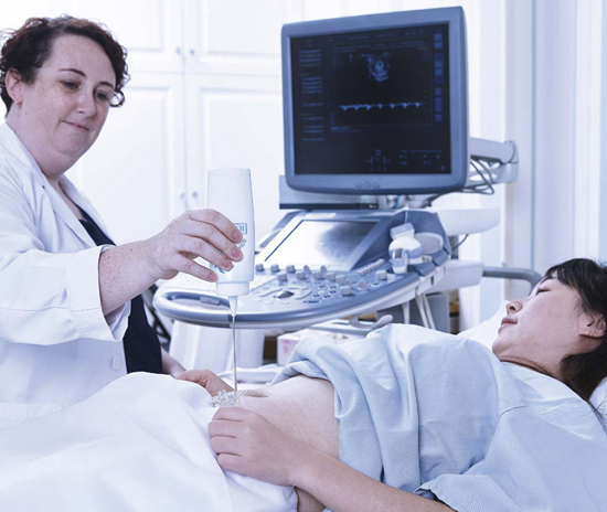
Ultrasound technicians apply ultrasound gel to the body to ensure a tight contact between the skin and the traducer, improving the transmission of sound waves.
The sound waves echo off the internal structures and are collected by the transducer as they reflect back. The returning sound waves are converted into images by the computer, which uses data such as the strength of the sound waves and the time it takes for them to reach the transducer. The result is the image that can be viewed on a screen in real time and recorded in forms such as still images or videos that may be printed out or stored as a digital file. The images produced by sonography are called sonograms, although they are sometimes referred to as ultrasounds as well.

