• Complain

Jacques S. Abramowicz - First-Trimester Ultrasound: A Comprehensive Guide

Here you can read online Jacques S. Abramowicz - First-Trimester Ultrasound: A Comprehensive Guide full text of the book (entire story) in english for free. Download pdf and epub, get meaning, cover and reviews about this ebook. year: 2015, publisher: Springer, genre: Romance novel. Description of the work, (preface) as well as reviews are available. Best literature library LitArk.com created for fans of good reading and offers a wide selection of genres:

Romance novel Science fiction Adventure Detective Science History Home and family Prose Art Politics Computer Non-fiction Religion Business Children Humor

Choose a favorite category and find really read worthwhile books. Enjoy immersion in the world of imagination, feel the emotions of the characters or learn something new for yourself, make an fascinating discovery.

Jacques S. Abramowicz First-Trimester Ultrasound: A Comprehensive Guide
  • Book:
    First-Trimester Ultrasound: A Comprehensive Guide
  • Author:
  • Publisher:
    Springer
  • Genre:
  • Year:
    2015
  • Rating:
    3 / 5
  • Favourites:
    Add to favourites
  • Your mark:
    • 60
    • 1
    • 2
    • 3
    • 4
    • 5

First-Trimester Ultrasound: A Comprehensive Guide: summary, description and annotation

We offer to read an annotation, description, summary or preface (depends on what the author of the book "First-Trimester Ultrasound: A Comprehensive Guide" wrote himself). If you haven't found the necessary information about the book — write in the comments, we will try to find it.

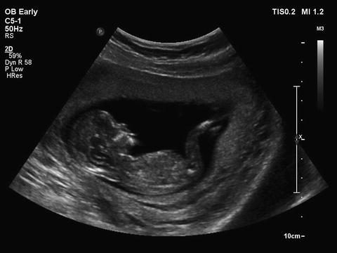
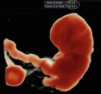
This book offers a unique and focused study of the use of ultrasound during the first trimester, a critical time in a fetus development. It includes basic examination guidelines as well as cutting-edge ultrasound modalities, including Doppler and three-dimensional ultrasound, for the period immediately preceding conception through early embryology. Beginning with a discussion of the safety and efficacy of diagnostic ultrasound and the use of this modality for the evaluation and treatment of infertility, recognized experts in the field explore conditions that may interfere with normal conception or development, including maternal diseases that would benefit from early scanning, elements of teratology, multiple gestations, ectopic pregnancy, gestational trophoblastic disease, fetal anomalies and invasive procedures in the first trimester. Numerous illustrations and figures are provided to serve as aids for understanding key concepts. First-Trimester Ultrasound is a valuable resource for many, in or after training, in obstetrics and gynecology, radiology, emergency medicine, family medicine and genetics.

Jacques S. Abramowicz: author's other books


Who wrote First-Trimester Ultrasound: A Comprehensive Guide? Find out the surname, the name of the author of the book and a list of all author's works by series.

First-Trimester Ultrasound: A Comprehensive Guide — read online for free the complete book (whole text) full work

Below is the text of the book, divided by pages. System saving the place of the last page read, allows you to conveniently read the book "First-Trimester Ultrasound: A Comprehensive Guide" online for free, without having to search again every time where you left off. Put a bookmark, and you can go to the page where you finished reading at any time.

Light

Font size:

Reset

Interval:

Bookmark:

Make
Springer International Publishing Switzerland 2016
Jacques S. Abramowicz (ed.) First-Trimester Ultrasound 10.1007/978-3-319-20203-7_1
1. Ultrasound in the First Trimester and Earlier: How to Keep It Safe
Jacques S. Abramowicz 1
(1)
Department of Obstetrics and Gynecology, Wayne State University School of Medicine, 3990 John R. Street, Detroit, MI 48201, USA
Jacques S. Abramowicz
Email:
Keywords
Artificial reproductive technologies (ART) Bioeffects Doppler Fetus First trimester Mechanical Index Pregnancy Risk Safety Teratology Thermal Index Ultrasound
Ultrasound is safe. Ultrasound is not X-rays. Our machines are FDA-approved. These are the statements most commonly made when initiating a conversation on safety of ultrasound. This is one of the reasons cited for its becoming an essential tool in medicine (together with its relatively low cost and immediate results availability). Diagnostic ultrasound (DUS) has been in use for over half a century in obstetrics and gynecology [].
Bioeffects of Ultrasound
Ultrasound is a waveform with a succession of positive and negative pressures [].
Thermal Effects
Human body normal core temperature is generally accepted to be 37 C with a diurnal variation of 0.51 C [].
Table 1.1
Changes over the years in I SPTA (in mW/cm2) in various medical applicationsa
Ultrasound clinical application
1976
1986
1991
Ophthalmic
Fetal, neonatal, pediatric imaging
Cardiac (adult)
Peripheral vascular
aAdapted from various sources []
Fig 11 In early pregnancy the entire fetus is within the ultrasound beam - photo 1
Fig. 1.1
In early pregnancy the entire fetus is within the ultrasound beam. Gestational age of 12 weeks
Non-thermal Effects
Ultrasound bioeffects may also occur through non-thermal or mechanical processes []. None of these have been demonstrated in humans and no harmful effects of diagnostic ultrasound, secondary to non-thermal mechanisms have been reported in human fetuses.
The Output Display Standard (OSD)
Until 1992 acoustic outputs of clinical ultrasound machines had specific limits. For instance, the upper limit of the spatial peak temporal average intensity or I SPTA (the most clinically useful intensity used to determine acoustic power of the ultrasound beam) for adult use was 720 mW/cm2 and for fetal use, 94 mW/cm2, which in fact, already had been increased from a previous maximum value of 46 mW/cm2. It was assumed that higher outputs would generate better images and, thus, improve diagnostic accuracy. Hence, end-users required ultrasound manufacturers to increase their machines output. Some worry, however, was expressed regarding the actual amount of energy absorbed by a human fetus during an ultrasound examination. This amount cannot be measured precisely. Not only the lack of an internal recording device is a major issue but, in addition, elements such as variations in maternal body habitus, fetal position changes, and gestational age progression render such a task impossible. To allow clinical users of ultrasound to use their instruments at higher powers than originally intended and to reflect the two major potential biological consequences of ultrasound (thermal and mechanical), the American Institute of Ultrasound in Medicine (AIUM), the National Electrical Manufacturers Association (NEMA) and the US food and Drug Administration (FDA), with representatives from the Canadian Health Protection Branch, the National Council on Radiation Protection and Measurements (NCRP) and 14 other medical organizations developed a standard related to the potential for ultrasound bioeffects []. The TI calculation is based on the formula:
First-Trimester Ultrasound A Comprehensive Guide - image 2
Fig. 1.2
The TI and MI acoustic indices as demonstrated on the monitor screen during routine ultrasound examination. In this picture, the MI is 0.9 and the TIS, 0.1
First-Trimester Ultrasound A Comprehensive Guide - image 3
where W is the acoustic power while scanning and W deg is the acoustic power required to achieve an increase in temperature of 1 C under similar conditions []. Furthermore, exposure time is not part of the equation, nor is it in the second index, the MI, which represents the potential for cavitation in tissues, but is not based on actual in situ measurements. The MI is defined as:
First-Trimester Ultrasound A Comprehensive Guide - image 4
It is a theoretical formulation of the ratio of the peak rarefaction pressure to the square root of the ultrasound frequency (hence, the higher the frequency, the lesser risk of mechanical effect, which is an advantage in endovaginal scanning). As for the TI, exposure time is not part of the calculation. Both the TI and MI can and should be followed as an indication of change in output during the clinical examination. A major component of the implementation of the ODs was supposed to be education of the end-user. Unfortunately, this aspect of the ODS does not seem to have succeeded as end-users knowledge of bioeffects, safety, and output indices is lacking. Both in Europe [].
Ultrasound and the Ovum
Ultrasound has permeated the field of infertility and reproductive endocrinology, from diagnosing uterine anomalies []. They wanted to determine how oocytes obtained under ultrasound guidance affected the pregnancy rate. The results obtained with 3.5-MHz probes suggest that exposure of human oocytes to ultrasonic waves during the different phases of meiosis does not significantly influence the developmental potential of the in vitro fertilized embryos. Unfortunately, no researcher describes any of the relevant exposure parameters discussed earlier, except for ultrasound frequency.
Ultrasound in Early Gestation
There are many valid medical indications to perform ultrasound in early gestation []. There was a timeeffect relation with activation of an enzyme pathway responsible for apoptosis through a mitochondrial pathway with exposures of 20 and 30 min but not 0 (control group) or 10 min.
Fetal Susceptibility to External Insults
The growing fetus is very sensitive to external influences. This is especially true in the first 1012 weeks of gestation []. If one, however, considers together the facts that hyperthermia is potentially harmful to the fetus and that ultrasound may, under certain circumstances elevate tissue temperature, then precaution has to be recommended, particularly in early gestation and especially with modes known to emit higher acoustic energy levels (such as pulsed Doppler).
Is Doppler Different and Can It Have Detrimental Effects on the Fetus in the First Trimester?
Ultrasound modalities can result in either scanned or unscanned exposure. Scanned conditions are associated with grey-scale B-mode images (the most commonly used real-time application), and Doppler images of tissue cross sections. Unscanned conditions are used for M-mode and pulsed-Doppler studies of tissue movement (such as cardiac valves) or blood velocity waveforms. This is clinically very important because for unscanned beams the power is limited to the area of the beam cross section, often very narrow (1 mm2) in the focal region. For scanned beams the acoustic power is not limited to a narrow area, but may cover large areas in the lateral direction, hence less risk of high exposure at a specific point. Furthermore, a variety of movements intervene during B-mode imaging, such as fetal body motion, observers hand movements, and maternal breathing. During a Doppler examination, however, it is necessary to have the transducer as steady as possible. This is because, in general, blood vessels or heart valves are small in comparison to the general organ or body size being scanned and even small movements will have more undesired effects on the resulting image. As described below, the most commonly used intensity (spatial peak temporal average intensity, I SPTA) associated with Doppler ultrasound is the highest of all the general-use categories, 1180 mW/cm2 for pulsed Doppler, as opposed to 34 mW/cm2 for B-mode, a 35-fold difference. Dwell time (duration of exposure) is also of major importance: Ziskin [].
Next page
Light

Font size:

Reset

Interval:

Bookmark:

Make

Similar books «First-Trimester Ultrasound: A Comprehensive Guide»

Look at similar books to First-Trimester Ultrasound: A Comprehensive Guide. We have selected literature similar in name and meaning in the hope of providing readers with more options to find new, interesting, not yet read works.


Reviews about «First-Trimester Ultrasound: A Comprehensive Guide»

Discussion, reviews of the book First-Trimester Ultrasound: A Comprehensive Guide and just readers' own opinions. Leave your comments, write what you think about the work, its meaning or the main characters. Specify what exactly you liked and what you didn't like, and why you think so.