Gebhard Mathis (editor) - Chest Sonography
Here you can read online Gebhard Mathis (editor) - Chest Sonography full text of the book (entire story) in english for free. Download pdf and epub, get meaning, cover and reviews about this ebook. year: 2022, publisher: Springer, genre: Romance novel. Description of the work, (preface) as well as reviews are available. Best literature library LitArk.com created for fans of good reading and offers a wide selection of genres:
Romance novel
Science fiction
Adventure
Detective
Science
History
Home and family
Prose
Art
Politics
Computer
Non-fiction
Religion
Business
Children
Humor
Choose a favorite category and find really read worthwhile books. Enjoy immersion in the world of imagination, feel the emotions of the characters or learn something new for yourself, make an fascinating discovery.

- Book:Chest Sonography
- Author:
- Publisher:Springer
- Genre:
- Year:2022
- Rating:5 / 5
- Favourites:Add to favourites
- Your mark:
Chest Sonography: summary, description and annotation
We offer to read an annotation, description, summary or preface (depends on what the author of the book "Chest Sonography" wrote himself). If you haven't found the necessary information about the book — write in the comments, we will try to find it.
This book, widely regarded as the standard work in the field, presents the state of the art in chest sonography, enhanced by a wealth of excellent illustrations. It provides the reader with concise, easy-to-assimilate information on all aspects of the use of the modality, including indications, investigative techniques, diagnostic decision making, and imaging artifacts and pitfalls. Chapters offer numerous tips and tricks and highlight potential diagnostic error sources to aid in daily clinical practice.
This sixth edition has been extensively revised to consider the latest techniques, study results, and meta-analyses and includes essential additional illustrative material. Chapter revisions include detailed guidance on contrast-enhanced ultrasound (CEUS) and the use of thoracic point-of-care ultrasound (PoCUS) in emergency patients.
As the techniques value and use continue to grow, readers will find Chest Sonography a valuable up-to-date resource and guide.
Gebhard Mathis (editor): author's other books
Who wrote Chest Sonography? Find out the surname, the name of the author of the book and a list of all author's works by series.


Chest Sonography — read online for free the complete book (whole text) full work
Below is the text of the book, divided by pages. System saving the place of the last page read, allows you to conveniently read the book "Chest Sonography" online for free, without having to search again every time where you left off. Put a bookmark, and you can go to the page where you finished reading at any time.
Font size:
Interval:
Bookmark:

This Springer imprint is published by the registered company Springer Nature Switzerland AG
The registered company address is: Gewerbestrasse 11, 6330 Cham, Switzerland
Ultrasonography of the lung has now become an established imaging procedure for chest diseases. Owing to comprehensive scientific investigations and studies (Beckh et al. ) the Point of Care Ultrasound (POCUS) has been widely accepted as a basic tool with rapid diagnostic information in medical examination.
In emergency cases and in intensive care medicine the findings of ultrasound allow a conclusive and strategic course of action (Diacon et al. ).
New developed handheld devices in the size of a mobile phone create an ultrasound-stethoscope. In this dimension ultrasound may provide an imaging diagnosis even in most difficult situations (Bachmann Nielsen et al. ).
The usefulness of chest sonography is proven in permanent enlargement of applications (Lesser ).
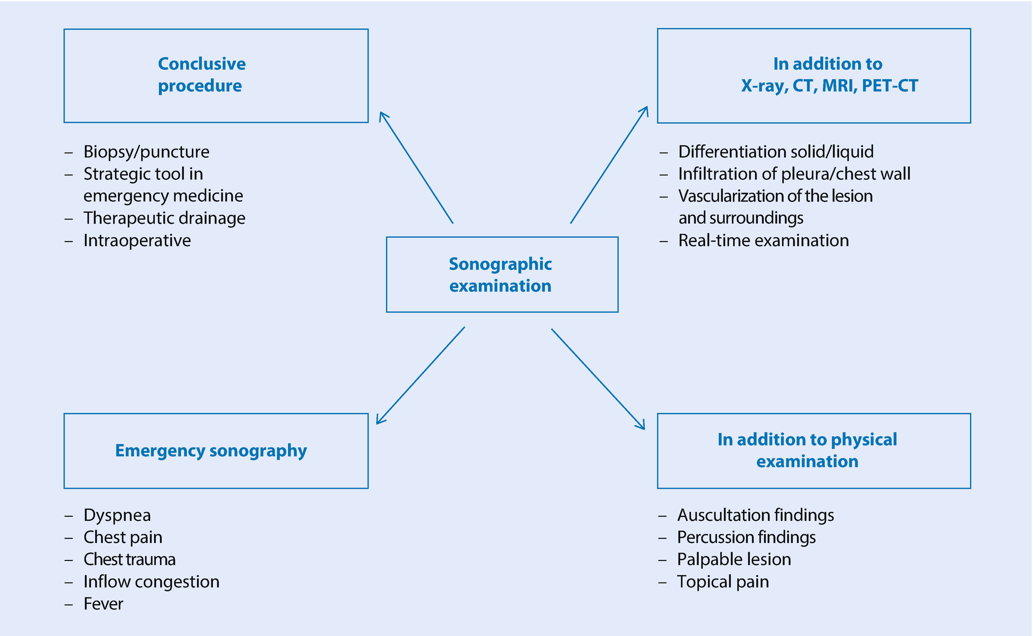
Spectrum of applications of ultrasonography for pleural and pulmonary diseases
Entities and pathological changes that can be accessed by ultrasound
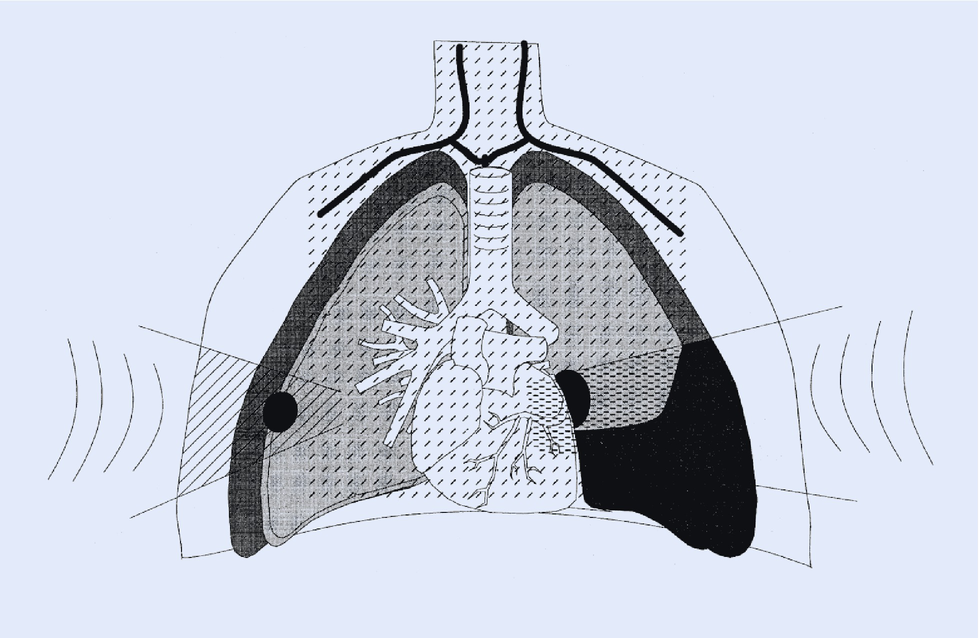
Acoustic shadowing occurs due to nearly complete absorption of the ultrasonic wave on bone, especially behind the sternum, the scapula, and the spine. Impairment due to the rib shadow can be balanced, at least in part, by appropriate breathing techniques.
The immediate retrosternal and posterior portions of the mediastinum cannot be viewed from the percutaneous aspect. Transesophageal and transbronchial ultrasound may be used additionally, but it should be noted that these examination procedures are invasive in terms of effort and handling (Lam and Becker ).
Ultrasonography provides diagnostic information during the investigation of individual entities in the chest (Overview).
- Chest wall
- Benign lesions:
Benign neoplasms (such as lipoma)
Hematoma
Abscess
Reactivated lymph nodes
Perichondritis, Tietze syndrome
Rib fracture
- Malignant lesions:
Lymph node metastases (primary diagnosis and the course of disease under treatment)
Growing invasive carcinomas
Osteolyses
- Pleura
- Solid structures:
Thickening of the pleura, callosity, calcification, asbestos-induced plaques
- Space-occupying lesion:
Benign: fibrous tumor, lipoma
Malignant: clearly identifiable metastases, diffuse carcinosis, pleural mesothelioma
- Fluid:
Effusion, hemothorax, pyothorax, chylothorax
- Dynamic investigation:
Pneumothorax
Differentiating between an effusion and a callosity
Adherence of a space-occupying lesion
Invasion by a space-occupying lesion
Mobility of the diaphragm
- Lung
Interstitial syndrome
- Benign peripheral lesions:
Inflammation, abscess, embolism, atelectasis
- Malignant peripheral lesions:
Peripheral metastasis, peripheral carcinoma, tumor/atelectasis
- Mediastinum, percutaneous:
Font size:
Interval:
Bookmark:
Similar books «Chest Sonography»
Look at similar books to Chest Sonography. We have selected literature similar in name and meaning in the hope of providing readers with more options to find new, interesting, not yet read works.
Discussion, reviews of the book Chest Sonography and just readers' own opinions. Leave your comments, write what you think about the work, its meaning or the main characters. Specify what exactly you liked and what you didn't like, and why you think so.