Part 1
Introduction
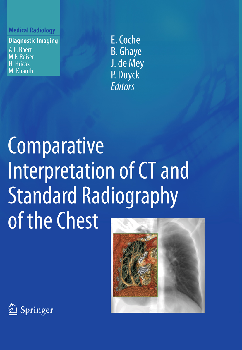
Emmanuel E. Coche , Benoit Ghaye , Johan de Mey and Philippe Duyck (eds.) Medical Radiology Comparative Interpretation of CT and Standard Radiography of the Chest 10.1007/978-3-540-79942-9_1 Springer Berlin Heidelberg 2011
1. Chest Radiography Today and Its Remaining Indications
Emmanuel E. Coche 1
(1)
Department of Medical Imaging, Cliniques Universitaires St-Luc, Avenue Hippocrate, 10, 1200 Brussels, Belgium
1.1
1.2
1.2.1
1.2.1.1
1.2.1.2
1.2.2
1.2.3
1.2.4
1.2.5
1.2.6
1.2.6.1
1.2.6.2
1.2.6.3
1.3
Abstract
Today, chest radiography remains the cornerstone for the diagnosis of many pulmonary diseases but it has received less attention during the past several years due to the explosion of new imaging techniques. However, chest radiography has many advantages. It offers simplicity, low cost, low radiation, large amounts of information, and is widely available throughout the world.
In this chapter, the remaining indications for the use of chest radiography in adults will be discussed in the context of MDCT. In pneumonia, chest radiography plays a pivotal role in the initial evaluation and follow-up of patients. In immunocompromised patients, chest radiography should be considered as a screening test and will often be followed by MDCT. In the intensive care unit (ICU), chest radiography can be easily performed to detect malposition of medical devices, but difficult cases still will be assessed by MDCT. In the emergency room, chest radiography is the recommended initial imaging study for the patient, and its results will often initiate additional imaging examinations such as MDCT. Chest radiography is also the most commonly used imaging tool for follow-up of benign conditions such as pneumothorax, pneumonia, pleural effusion. Its role in the follow-up of malignant conditions is less obvious. At the end of the chapter, the role of chest radiography in lung cancer screening, assessment of preoperative patients, and daily follow-up will be discussed.
1.1 Introduction
Despite continuous advances in cross-sectional imaging, chest radiography remains the cornerstone for the diagnosis of many pulmonary diseases and the most commonly performed diagnostic imaging test in western countries and probably throughout the world. This imaging modality is responsible for approximately 3040% of all X-ray examinations performed, regardless of the level of health care delivery. International commission on radiological protection (ICRP 1993). Over the period of 19931999, chest radiography was the most frequently used radiologic examination in the United States (Maitino et al. ).
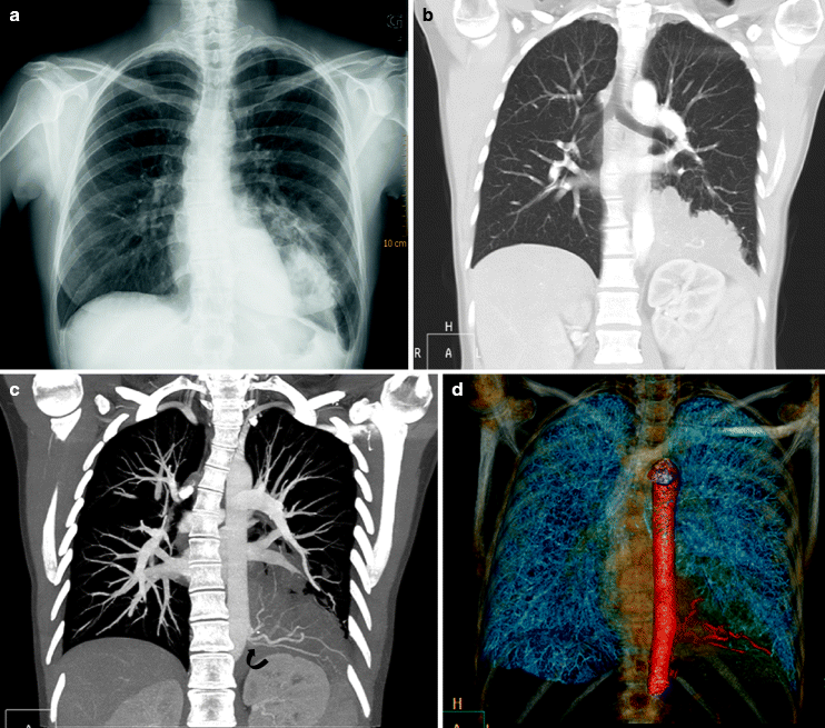
Fig. 1.1
Images obtained from a 35-year-old woman referred for chronic cough and fever. ( a ) Chest radiography demonstrates a left retrocardiac mass containing a lucent area. ( b ) Frontal reformatted CT image demonstrates, in the lung window setting, the irregular borders of the mass. ( c ) Maximal Intensity Projection (MIP) image demonstrates the presence of aberrant vessels ( curved arrow ) originating directly from the thoracic aorta and feeding the lung abnormality. ( d ) Volume rendered image highlights in red the aberrant vessels linked to an intralobar sequestration
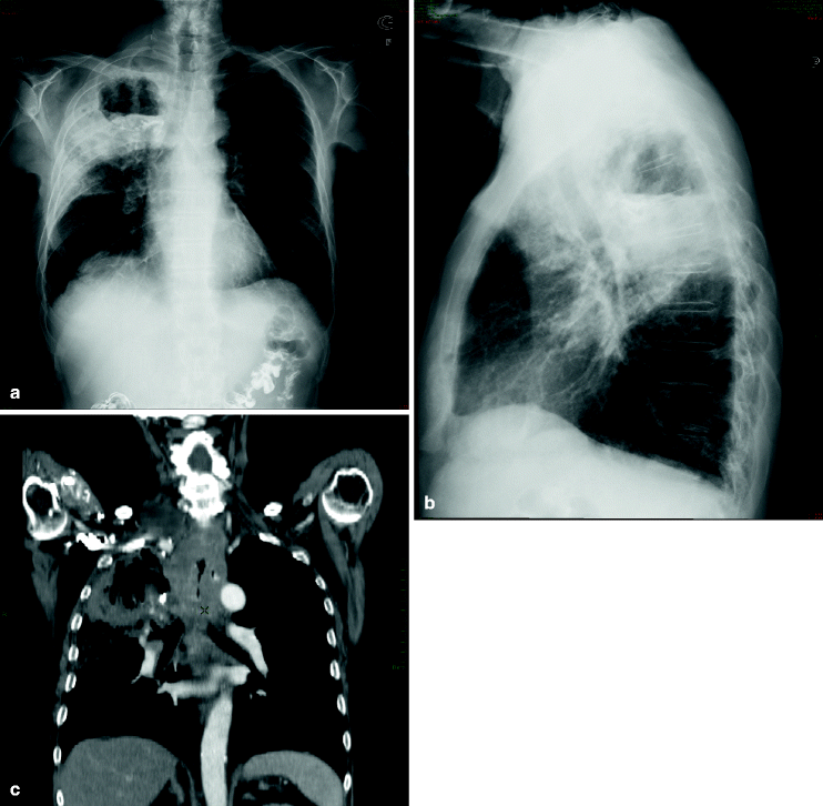
Fig. 1.2
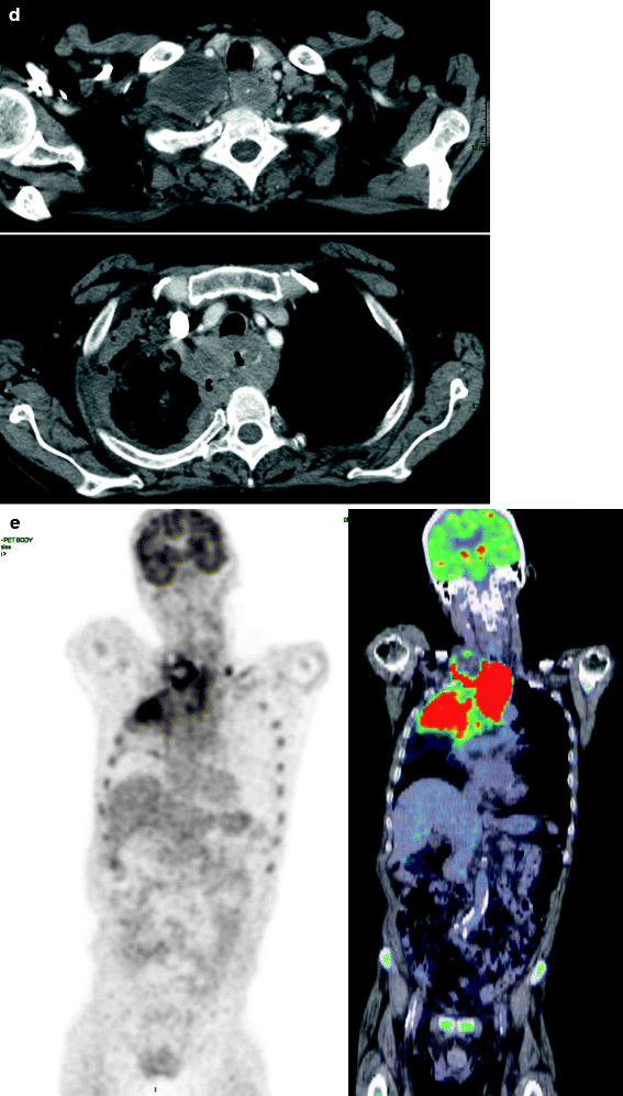
Images obtained from a 76-year-old man with previous oropharyngeal carcinoma addressed for dysphagia and weight loss since 1 month. ( a ) Posteroanterior chest radiography reveals a large density of the right upper thorax containing a fluid level. ( b ) Lateral chest radiograph shows the posterior location of this abnormality. ( c ) Frontal reformation of spiral CT acquisition on mediastinal window setting reveals soft-tissue infiltration of the mediastinum centered on the esophagus and a large aerated cavity in the right upper lobe. ( d ) Two axial CT scans obtained at the level of the upper chest confirm the esophageal thickening. Note the right pleural effusion. ( e ) 18Fludeoxyglucose Positron emission tomography with frontal reformation reveals intense uptake in the area of the mediastinal thickening and the right upper lobe. ( f ) Barium swallowing demonstrates a large fistula between the upper esophagus and the right lung. ( g ) Axial CT scan performed after oral opacification confirms the abnormal communication between the esophagus and the right lung. Esophageal biopsy was performed, and pathology revealed a well-differentiated squamous cell carcinoma of the esophagus
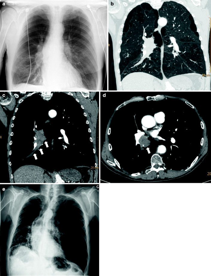
Fig. 1.3
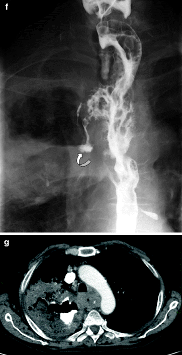
Images obtained from a 75-year-old man with chronic obstructive pulmonary disease admitted to the emergency department for exacerbation of dyspnea. ( a ) At admission, anteroposterior chest radiograph does not show any obvious abnormality. ( b ) Enhanced spiral MDCT was performed to rule out pulmonary embolism. Frontal reformation demonstrates on the lung window setting a large right hilar mass with right lower lobe mucous plugging. Note the paraseptal emphysema in both upper lobes. ( c ) The mediastinal window setting depicts well the right hilar and bronchial abnormalities ( straight arrows ). ( d ) Axial CT at the level of the aortic root shows a large right hilar ( straight arrow ) mass in relation to a squamous cell carcinoma (proved pathologically). ( e ) Chest radiograph performed several days after admission shows a mediastinal shift to the right linked to a complete obstruction of the right inferior bronchus and lung collapse.This case illustrates the usefulness of MDCT in case of discordant results between chest radiography and the patients complaints
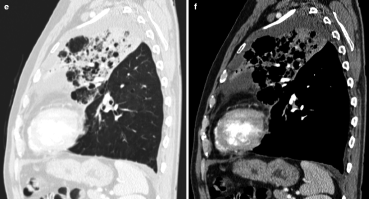
Fig. 1.4
Images obtained from a 65-year-old man who was admitted for fever and dyspnea. ( a ) Postero-anterior osteroanterior chest radiograph shows a left upper lobe consolidation with air bronchogram suggestive of lobar pneumonia. ( b ) Lateral chest radiography confirms the consolidation delimited by the great fissure posteriorly and involving the left upper lobe. Multiple small radiolucencies are observed within the consolidation area. ( c ) Frontal reformation of spiral CT acquisition on lung window setting confirms the infectious nature of the left upper lobe consolidation. Note the nice depiction of small radiolucencies linked to preexisting emphysema. ( d ) Frontal reformation of spiral CT acquisition on mediastinal window setting rules out the possibility of an obstructive lung carcinoma. ( e ) Sagittal reformation of spiral CT acquisition on lung window setting shows the nice side-by-side comparison with the corresponding chest radiography. ( f ) Sagittal reformation of spiral CT acquisition on mediastinal window setting highlights the anatomical delineation of the consolidation by the great fissure