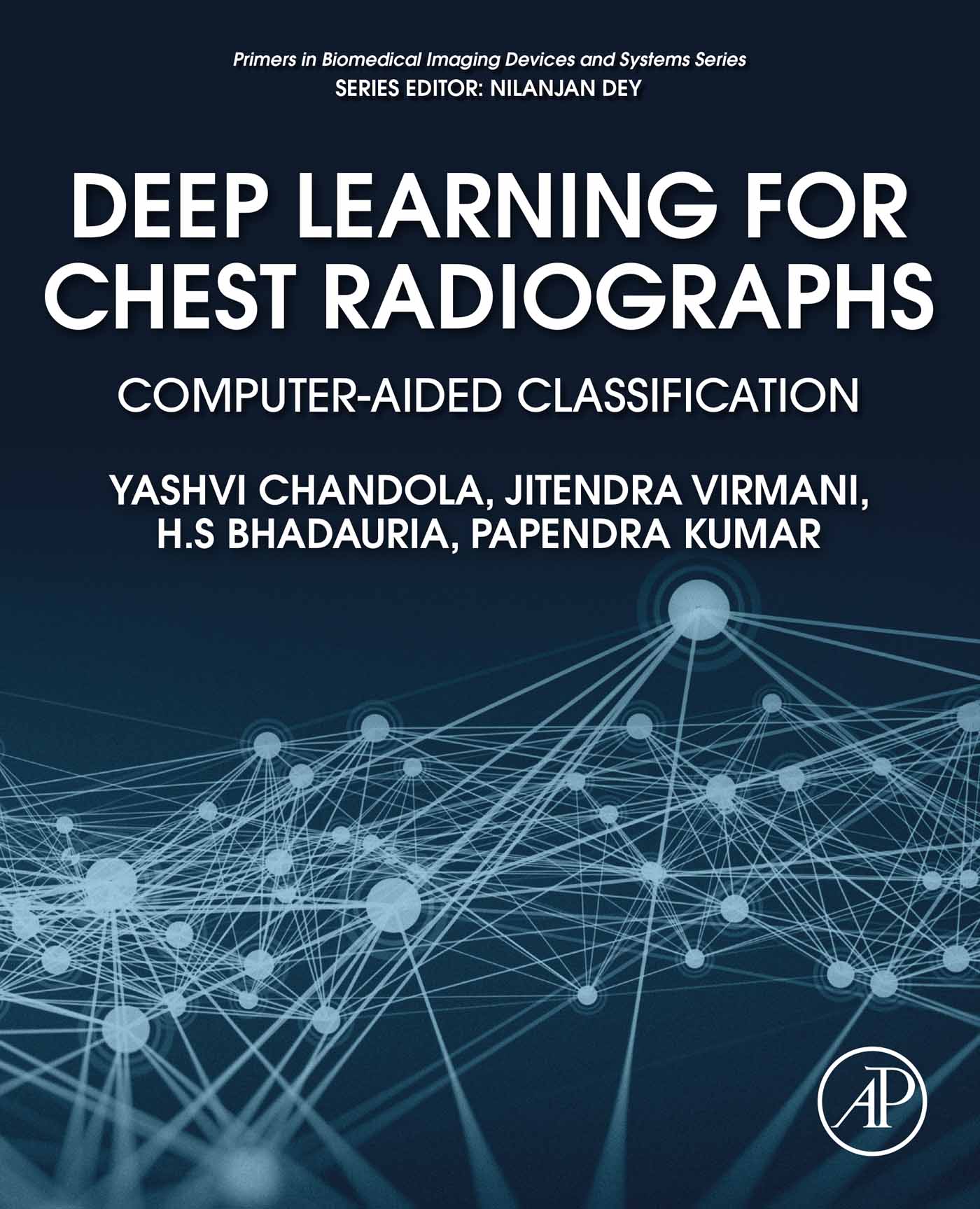
Yashvi Chandola - Deep Learning for Chest Radiographs: Computer-Aided Classification (Primers in Biomedical Imaging Devices and Systems)
Here you can read online Yashvi Chandola - Deep Learning for Chest Radiographs: Computer-Aided Classification (Primers in Biomedical Imaging Devices and Systems) full text of the book (entire story) in english for free. Download pdf and epub, get meaning, cover and reviews about this ebook. year: 2021, publisher: Academic Press, genre: Romance novel. Description of the work, (preface) as well as reviews are available. Best literature library LitArk.com created for fans of good reading and offers a wide selection of genres:
Romance novel
Science fiction
Adventure
Detective
Science
History
Home and family
Prose
Art
Politics
Computer
Non-fiction
Religion
Business
Children
Humor
Choose a favorite category and find really read worthwhile books. Enjoy immersion in the world of imagination, feel the emotions of the characters or learn something new for yourself, make an fascinating discovery.

- Book:Deep Learning for Chest Radiographs: Computer-Aided Classification (Primers in Biomedical Imaging Devices and Systems)
- Author:
- Publisher:Academic Press
- Genre:
- Year:2021
- Rating:5 / 5
- Favourites:Add to favourites
- Your mark:
Deep Learning for Chest Radiographs: Computer-Aided Classification (Primers in Biomedical Imaging Devices and Systems): summary, description and annotation
We offer to read an annotation, description, summary or preface (depends on what the author of the book "Deep Learning for Chest Radiographs: Computer-Aided Classification (Primers in Biomedical Imaging Devices and Systems)" wrote himself). If you haven't found the necessary information about the book — write in the comments, we will try to find it.
Deep Learning for Chest Radiographs enumerates different strategies implemented by the authors for designing an efficient convolution neural network-based computer-aided classification (CAC) system for binary classification of chest radiographs into Normal and Pneumonia. Pneumonia is an infectious disease mostly caused by a bacteria or a virus. The prime targets of this infectious disease are children below the age of 5 and adults above the age of 65, mostly due to their poor immunity and lower rates of recovery. Globally, pneumonia has prevalent footprints and kills more children as compared to any other immunity-based disease, causing up to 15% of child deaths per year, especially in developing countries. Out of all the available imaging modalities, such as computed tomography, radiography or X-ray, magnetic resonance imaging, ultrasound, and so on, chest radiographs are most widely used for differential diagnosis between Normal and Pneumonia. In the CAC system designs implemented in this book, a total of 200 chest radiograph images consisting of 100 Normal images and 100 Pneumonia images have been used. These chest radiographs are augmented using geometric transformations, such as rotation, translation, and flipping, to increase the size of the dataset for efficient training of the Convolutional Neural Networks (CNNs). A total of 12 experiments were conducted for the binary classification of chest radiographs into Normal and Pneumonia. It also includes in-depth implementation strategies of exhaustive experimentation carried out using transfer learning-based approaches with decision fusion, deep feature extraction, feature selection, feature dimensionality reduction, and machine learning-based classifiers for implementation of end-to-end CNN-based CAC system designs, lightweight CNN-based CAC system designs, and hybrid CAC system designs for chest radiographs.
This book is a valuable resource for academicians, researchers, clinicians, postgraduate and graduate students in medical imaging, CAC, computer-aided diagnosis, computer science and engineering, electrical and electronics engineering, biomedical engineering, bioinformatics, bioengineering, and professionals from the IT industry.
Yashvi Chandola: author's other books
Who wrote Deep Learning for Chest Radiographs: Computer-Aided Classification (Primers in Biomedical Imaging Devices and Systems)? Find out the surname, the name of the author of the book and a list of all author's works by series.

Deep Learning for Chest Radiographs: Computer-Aided Classification (Primers in Biomedical Imaging Devices and Systems) — read online for free the complete book (whole text) full work
Below is the text of the book, divided by pages. System saving the place of the last page read, allows you to conveniently read the book "Deep Learning for Chest Radiographs: Computer-Aided Classification (Primers in Biomedical Imaging Devices and Systems)" online for free, without having to search again every time where you left off. Put a bookmark, and you can go to the page where you finished reading at any time.
Font size:
Interval:
Bookmark:
Academic Press is an imprint of Elsevier
125 London Wall, London EC2Y 5AS, United Kingdom
525 B Street, Suite 1650, San Diego, CA 92101, United States
50 Hampshire Street, 5th Floor, Cambridge, MA 02139, United States
The Boulevard, Langford Lane, Kidlington, Oxford OX5 1GB, United Kingdom
Copyright 2021 Elsevier Inc. All rights reserved.
No part of this publication may be reproduced or transmitted in any form or by any means, electronic or mechanical, including photocopying, recording, or any information storage and retrieval system, without permission in writing from the publisher. Details on how to seek permission, further information about the Publishers permissions policies and our arrangements with organizations such as the Copyright Clearance Center and the Copyright Licensing Agency, can be found at our website: www.elsevier.com/permissions.
This book and the individual contributions contained in it are protected under copyright by the Publisher (other than as may be noted herein).
Notices
Knowledge and best practice in this field are constantly changing. As new research and experience broaden our understanding, changes in research methods, professional practices, or medical treatment may become necessary.
Practitioners and researchers must always rely on their own experience and knowledge in evaluating and using any information, methods, compounds, or experiments described herein. In using such information or methods they should be mindful of their own safety and the safety of others, including parties for whom they have a professional responsibility.
To the fullest extent of the law, neither the Publisher nor the authors, contributors, or editors, assume any liability for any injury and/or damage to persons or property as a matter of products liability, negligence or otherwise, or from any use or operation of any methods, products, instructions, or ideas contained in the material herein.
Library of Congress Cataloging-in-Publication Data
A catalog record for this book is available from the Library of Congress
British Library Cataloguing-in-Publication Data
A catalogue record for this book is available from the British Library
ISBN 978-0-323-90184-0
For information on all Academic Press publications visit our website at https://www.elsevier.com/books-and-journals

Publisher: Mara Conner
Acquisitions Editor: Sonnini Yura
Editorial Project Manager: Leticia Lima
Production Project Manager: Poulouse Joseph
Cover Designer: Mark Rogers
Typeset by SPi Global, India
Yashvi Chandola; Jitendra Virmani; H.S. Bhadauria; Papendra Kumar
Medical images are an irreplaceable source of statistical information for the purpose of extensive scientific experimental applications. The imaging modalities that facilitate the capturing of these medical images are evolving speedily, thereby improvising the sophistication in the quality of images. This affects the features associated with different tissues at reasonably subsiding costs. Consequently, in this era of data-driven disciplines, the medical images result in a noteworthy increase in the repository size of the image data. As medical imaging datasets grow grander and become more complicated, the need arises for more advanced machine learning-based methods to analyze and interpret the data. During the past few years, deep learning-based methods have brought a revolution to the field of medical image analysis by introducing novel, efficient solutions to many image analysis problems. These problems are as diverse as computer-aided classification, segmentation of regions of interest, lesion detection, image-guided therapy, and image dataset annotation. Machine learning and deep learning provide a wide range of tools and methods to execute the solutions of this diverse pool of problems.
The principal aim of Deep Learning for Chest Radiographs: Computer-Assisted Classification is to describe in detail: (a) the different types of computer-assisted classification systems for chest radiographs; (b) the design of computer-aided classification systems for chest radiographs using end-to-end convolution neural networks; (c) the design of computer-aided classification systems for chest radiographs using end-to-end lightweight convolution neural networks; and (d) the design of computer-aided classification systems for chest radiographs using hybrid approaches like deep feature extraction by convolutional neural network and classification using conventional machine learning classifiers. The authors have created this volume keeping in mind a diverse readership, but the prime target audience are the graduates, undergraduates, academic researchers, research scholars, and industry personnel who are fascinated by the trends in the application of these machine learning and deep learning methods for the design of computer-aided classification systems for medical images in general and chest radiographs in particular. The organization of the book keeps in mind the majority of the readership audience, hence the chapters summarize the basic concepts of deep learning-based convolution neural network-based architectures, machine learning algorithms in medical imaging with prime focus on chest radiographs, the various data augmentation methods, features of medical images that are essential in correct diagnosis of diseases. Each chapter has a set of code snippets as well as illustrations of how to apply these methods focusing on chest radiographs. The topics discussed in this book mirror the persistent progress of deep learning approaches for the analysis of medical images. The authors hope that this book will help the readers understand the current research in medical image analysis and opportunities for developing pioneering computational methods, which will positively accelerate the discoveries of computer-aided classification systems into effective treatments for patients. The authors of this book share the common vision of achieving greater advancements in the field of medical image analysis and are grateful for the opportunity to contribute their knowledge, aptitude, and familiarity with the topic.
Yashvi Chandola; Jitendra Virmani; H.S. Bhadauria; Papendra Kumar
This book has been one of the significant academic achievements for all of the contributing authors and has been possible by their combined efforts. We have had the utmost honor and privilege of working with a very professional team at Elsevier, and we are thankful for their timely guidance and support from the initial review of the book proposal to the final copy editing stage and production. With the greatest regards, we acknowledge our heartfelt and sincere thanks to Dr. Nilanjan Dey (series editor) for the critical review of the proposal and for rendering his valuable advice from time to time. The authors are also thankful to Dr. Kriti and Mr. Niranjan Yadav for their support and help in stimulating discussions on different topics covered in this book. The authors are grateful to Mr. V.M. Thakkar, Mr. Bhoopender Chandola, Dr. Jyoti Rawat, and Mrs. Suman Lata Joshi for their constructive suggestions and help rendered in proofreading the manuscript. We extend our utmost gratitude to the participating radiation oncologist Dr. Yamini Bachheti (M.B.B.S, M.D. Radiation Oncology), currently serving as a senior resident at All India Institute of Medical Sciences, Rishikesh, for her valuable inputs during the numerous discussions we had throughout the progress of the work. She made us understand how the differential diagnosis between different classes of chest radiographs is carried out by visual analysis.
Font size:
Interval:
Bookmark:
Similar books «Deep Learning for Chest Radiographs: Computer-Aided Classification (Primers in Biomedical Imaging Devices and Systems)»
Look at similar books to Deep Learning for Chest Radiographs: Computer-Aided Classification (Primers in Biomedical Imaging Devices and Systems). We have selected literature similar in name and meaning in the hope of providing readers with more options to find new, interesting, not yet read works.
Discussion, reviews of the book Deep Learning for Chest Radiographs: Computer-Aided Classification (Primers in Biomedical Imaging Devices and Systems) and just readers' own opinions. Leave your comments, write what you think about the work, its meaning or the main characters. Specify what exactly you liked and what you didn't like, and why you think so.