Table of Contents
List of tables
- Tables in Chapter1
- Tables in Chapter2
- Tables in Chapter3
- Tables in Chapter6
- Tables in Chapter7
- Tables in Chapter8
- Tables in Chapter9
- Tables in Chapter11
- Tables in Chapter12
- Tables in Chapter13
- Tables in Chapter14
- Tables in Chapter15
- Tables in Chapter16
- Tables in Chapter17
- Tables in Chapter18
- Tables in Chapter19
- Tables in Chapter20
- Tables in Chapter21
- Tables in Chapter22
- Tables in Chapter23
- Tables in Chapter24
- Tables in Chapter25
- Tables in Chapter26
- Tables in Chapter27
- Tables in Chapter28
- Tables in Nuclear Medicine
- Tables in The ABCs of Heart Disease
List of figures
- Figures in Chapter1
- Figures in Chapter2
- Figures in Chapter3
- Figures in Chapter4
- Figures in Chapter5
- Figures in Chapter6
- Figures in Chapter7
- Figures in Chapter8
- Figures in Chapter9
- Figures in Chapter10
- Figures in Chapter11
- Figures in Chapter12
- Figures in Chapter13
- Figures in Chapter14
- Figures in Chapter15
- Figures in Chapter16
- Figures in Chapter17
- Figures in Chapter18
- Figures in Chapter19
- Figures in Chapter20
- Figures in Chapter21
- Figures in Chapter22
- Figures in Chapter23
- Figures in Chapter24
- Figures in Chapter25
- Figures in Chapter26
- Figures in Chapter27
- Figures in Chapter28
- Figures in Nuclear Medicine
- Figures in The ABCs of Heart Disease
- Figures in Chapter 1 Quiz Answers
- Figures in Appendix
Landmarks
Acknowledgments
I am again grateful to the many thousands of you whom I have never met but who found a website called Learning Radiology helpful, making it so popular that it played a role launching the first edition of this book, which itself was so popular that it led to this third edition.
For their help and suggestions, I thank David Saul, MD, one of our radiology residents, who made invaluable suggestions about how this edition could be changed. Daniel Kowal, MD, a radiologist who graduated from our program, did an absolutely wonderful job in simplifying the complexities of MRI again in the chapter he wrote. Jeffrey Cruz, MD, one of our residents, helped out with the online Radiation Safety and Dose module, and Sherif Saad, MD, contributed an illustration.
I thank Chris Kim, MD; Susan Summerton, MD; Mindy Horrow, MD; Peter Wang, MD; and Huyen Tran, MD, for supplying additional images for this edition. And thanks to Mindy Horrow, MD; Eric Faerber, MD; and Brooke Devenney-Cakir, MD, for reviewing chapters from this text.
I certainly want to recognize and again thank Jim Merritt and Katy Meert from Elsevier for their support and assistance.
I also acknowledge the hundreds of radiology residents and medical students who, over the years, have provided me with an audience of motivated learners, without whom a teacher would have no one to teach.
Finally, I want to thank my wonderful wife, Pat, who has encouraged me throughout the project, and my family.
William Herring MD, FACR
Appendix
What to order when
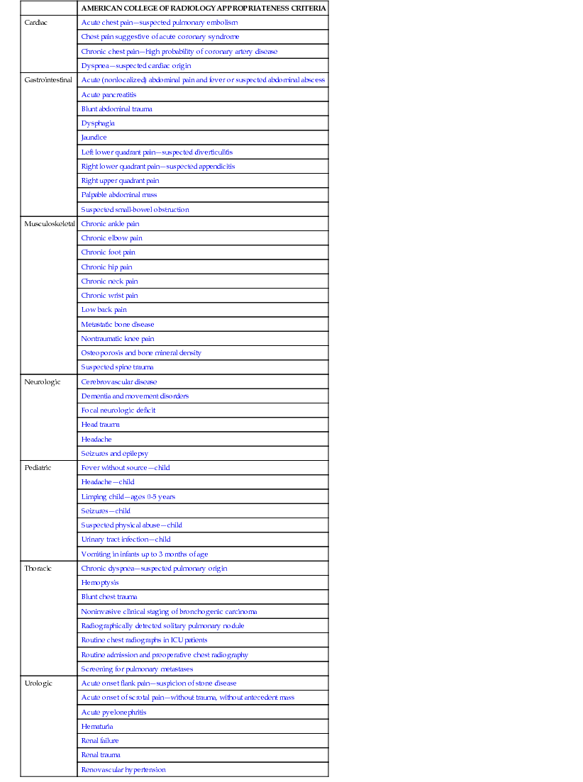
The links to the American College of Radiology's Appropriateness Criteria provided below explain which imaging study to order under certain clinical circumstances. These guidelines were developed by a series of expert panels consisting of diagnostic radiologists, interventional radiologists, and radiation oncologists, as well as leaders in other specialties. These are evidence-based guidelines designed to assist health-care providers in making the most appropriate imaging or treatment decision for a patient with a specific clinical condition.
| AMERICAN COLLEGE OF RADIOLOGY APPROPRIATENESS CRITERIA |
| Cardiac | Acute chest painsuspected pulmonary embolism |
| Chest pain suggestive of acute coronary syndrome |
| Chronic chest painhigh probability of coronary artery disease |
| Dyspneasuspected cardiac origin |
| Gastrointestinal | Acute (nonlocalized) abdominal pain and fever or suspected abdominal abscess |
| Acute pancreatitis |
| Blunt abdominal trauma |
| Dysphagia |
| Jaundice |
| Left lower quadrant painsuspected diverticulitis |
| Right lower quadrant painsuspected appendicitis |
| Right upper quadrant pain |
| Palpable abdominal mass |
| Suspected small-bowel obstruction |
| Musculoskeletal | Chronic ankle pain |
| Chronic elbow pain |
| Chronic foot pain |
| Chronic hip pain |
| Chronic neck pain |
| Chronic wrist pain |
| Low back pain |
| Metastatic bone disease |
| Nontraumatic knee pain |
| Osteoporosis and bone mineral density |
| Suspected spine trauma |
| Neurologic | Cerebrovascular disease |
| Dementia and movement disorders |
| Focal neurologic deficit |
| Head trauma |
| Headache |
| Seizures and epilepsy |
| Pediatric | Fever without sourcechild |
| Headachechild |
| Limping childages 0-5 years |
| Seizureschild |
| Suspected physical abusechild |
| Urinary tract infectionchild |
| Vomiting in infants up to 3 months of age |
| Thoracic | Chronic dyspneasuspected pulmonary origin |
| Hemoptysis |
| Blunt chest trauma |
| Noninvasive clinical staging of bronchogenic carcinoma |
| Radiographically detected solitary pulmonary nodule |
| Routine chest radiographs in ICU patients |
| Routine admission and preoperative chest radiography |
| Screening for pulmonary metastases |
| Urologic | Acute onset flank painsuspicion of stone disease |
| Acute onset of scrotal painwithout trauma, without antecedent mass |
| Acute pyelonephritis |
| Hematuria |
| Renal failure |
| Renal trauma |
| Renovascular hypertension |
Chapter 1 Quiz Answers
Here you are at the end of the book. Finished the text already? That was speedy reading on your part. Here are the answers to the quiz that appears in .
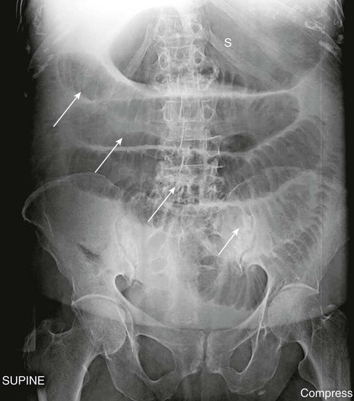
FIGURE 1-1 Small-bowel obstruction. There are multiple air-filled and dilated loops of small bowel (white arrows) with virtually no gas in the large bowel. The stomach (S) is also dilated. The disproportionate dilatation of small bowel is indicative of a mechanical small bowel obstruction caused, in this case, by adhesions from previous surgery.
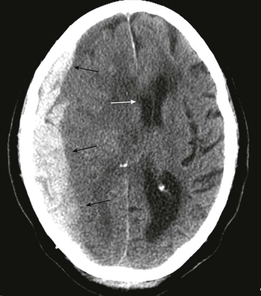
FIGURE 1-2 Subdural hematoma. A curvilinear band of increased attenuation in the right parietal region (black arrows) is causing a subfalcine shift of the midline structures to the left (white arrow). The crescentric increased density, paralleling the inner table, is classic for a subdural collection. The patient fell from a height and struck his head.


![William Herring - Learning Radiology: Recognizing the Basics [with Student Consult Online Access]](/uploads/posts/book/310283/thumbs/william-herring-learning-radiology-recognizing.jpg)
