Springer-Verlag Berlin Heidelberg 2016
Seung-Kyu Han Innovations and Advances in Wound Healing 10.1007/978-3-662-46587-5_1
1. Basics of Wound Healing
Seung-Kyu Han 1
(1)
Professor of Plastic Surgery, Director of Diabetic Wound Center, Director of Cell Therapy Laboratory, Korea University College of Medicine and Korea University Guro Hospital, Seoul, Republic of Korea (South Korea)
Abstract
We all experience our fair share of wounds during the course of our lives. We get pricked by thorns, scratched by sharp objects, sunburned at the beach, and scalded by hot water. Accidents can lead to our skin being peeled or sliced off, and many of us may undergo surgical procedures which inevitably result in surgical wounds on our bodies. Selecting an appropriate wound healing strategy is crucial for successful wound healing in that it can minimize the risk of complications, enhance the speed of wound healing, and minimize scar formation after the wound has fully healed. During the past few decades, various technologies have been developed for optimal wound healing. In order to understand new techniques, procedure, and materials in wound healing, medical professionals should have a basic knowledge of wound healing. In this chapter, clinically useful anatomy of the skin, terminology and documentation for wounds, basic wound healing process, and conventional wound healing methods will be briefly described prior to the main topics of this book.
Keywords
Anatomy Skin Wound healing
Clinical Anatomy of the Skin
Our skin layer has many crucial functions, but the main role is barrier function, that is, protecting our body from external elements. Unless the wound is properly healed, the integrity of the skin barrier is compromised and allows external germs such as bacteria and viruses to freely invade our body. This can even lead to critical organ damage. Moreover, the skin layer comprises an important aspect of ones appearance, which of course has a significant influence on ones social life; thus, the skin layer can also be regarded as an important factor of mental health.
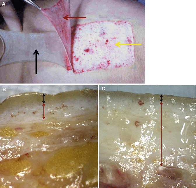
Since the skin is such a critical element of both physical and mental health, optimal care must be given to any sustained wounds, so that the damaged skin region is restored to a structure and form similar to the original as quickly as possible for the skin to resume its functions. In order to give such optimal wound care, one must first understand the anatomy of the skin. The skin is the largest organ of the body. It weighs in the range of 2.73.6 kg and receives 1/3 of the bodys blood volume. The thickness of the skin varies from 0.5 to 6.0 mm. The skin consists of cells and extracellular matrices. There are three layers in the skin. The epidermis is the thin, outer layer of the skin. The dermis is a thicker, inner layer. The subcutaneous fatty tissue (hypodermis) is a layer of loose connective tissue lying beneath the dermis (Fig. ).
Fig. 1.1
Skin layers. ( A ) Epidermis ( a black arrow ), dermis ( a red arrow ), and hypodermis ( a yellow arrow ). ( B ) Epidermis ( a black arrow ) and dermis ( a red arrow ) of the thin skin (hand dorsum). ( C ) Epidermis ( a black arrow ) and dermis ( a red arrow ) of the thick skin (palm skin)
Epidermis
The thickness of the epidermis varies in different types of skin. It is the thinnest on the eyelids at 0.05 mm and the thickest on the palms and soles at 1.5 mm. The epidermis is an avascular layer receiving blood supply from the dermis across the semipermeable basement membrane.
Epidermal Layers
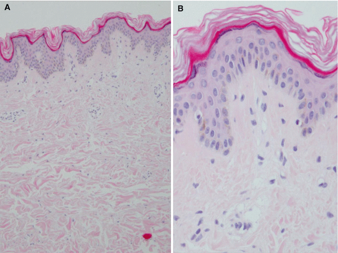
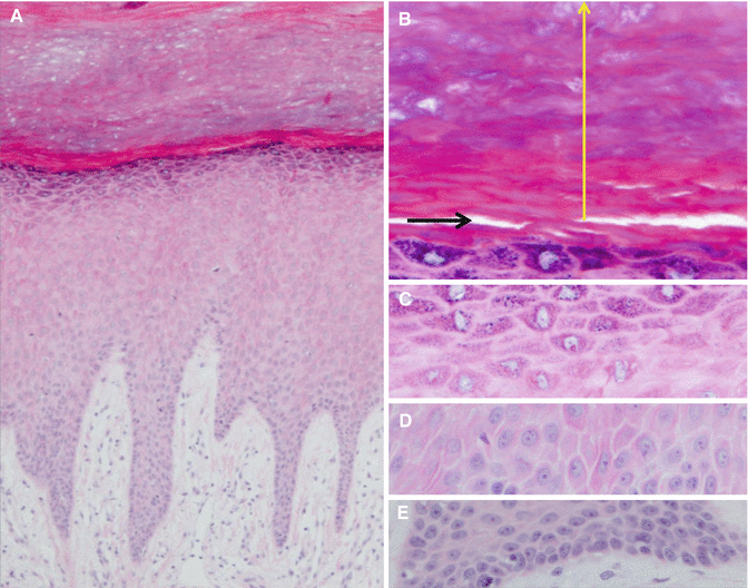
The epidermis contains five layers. From bottom to top, the layers are named stratum basale, stratum spinosum, stratum granulosum, stratum lucidum, and stratum corneum. The thickness and layers of the epidermis vary depending on the location of the body (Figs. ).
Fig. 1.2
Epidermis of the thigh. The number of epidermal layer is small. ( A ) 100. ( B ) 400
Fig. 1.3
Epidermis of the plantar skin. In comparison with the thigh skin (Fig. ), there are much more layers in the epidermis. ( A ) 100. ( B ) 400. Horny ( a yellow arrow ) and lucid ( a black arrow ) layers. ( C ) Granular layer. 400. ( D ) Spinous layer. 400. ( E ) Basal layer. 400
The stratum basale (basal layer) is the deepest layer with a thickness of a single cell. It is the only layer of the epidermis in which cells undergo mitosis. The stratum basale forms the dermal-epidermal junction (basement membrane zone), which separates the epidermis from the dermis. The stratum spinosum (spinous layer) consists of several rows of more mature keratinocytes, which appear spiny under a microscope. The stratum granulosum (granular layer) contains 35 flattened cell rows comprising a higher concentration of keratin. The stratum lucidum (lucid layer) is a thin, clear layer of dead skin cells found in the thick skin such as the palms and soles. The stratum corneum (horny layer) consists of dead cells (corneocytes) and keratin. This layer prevents water evaporation, absorbs water, and easily sheds itself.
Epidermal Cells
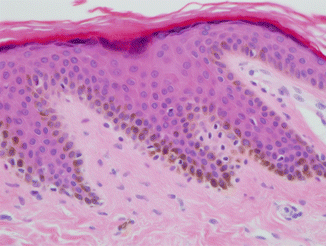
The keratinocytes are major cells of the epidermis, making up approximately 90 % of epidermal cells. These cells are responsible for the toughness of the skin. They produce keratin and form the basic component of hair, skin, and nails. Langerhans cells protect the body against infection by attacking and engulfing foreign materials. Melanocytes are responsible for producing melanin (Fig. ). While the number of melanocytes remains the same in individual body regions in all human beings, their activity can vary between different body regions and across individuals. In white and oriental skin, the melanosomes are packed in aggregates, but in black skin, they are larger and distributed more evenly. The number of melanosomes in the keratinocytes increases with ultraviolet (UV) radiation exposure, while their distribution remains largely unaffected. Merkel cells are mechanoreceptors that provide information on light touch sensation.
Fig. 1.4
Melanocytes and melanin ( brown color )
Epidermal Appendages
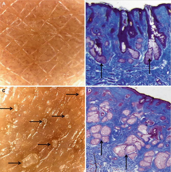
The sebaceous glands secrete sebum into hair follicles. Sebum is an oily substance, which provides moisture and softness to the skin. Sebum also acts as a barrier against foreign substances (Fig. ). Hair contributes to the appearance, body temperature, protection, and sensation. The eccrine sweat glands produce sweat to help regulate body temperature and assist with elimination of waste products. The apocrine sweat glands produce sweat, which is responsible for body odor. Nails are made of dead cells that contain keratin.
Fig. 1.5
Differences can be observed in the development of the sebaceous glands according to each individual. ( A , B ) Skin with less developed sebaceous glands ( arrows ). ( C , D ) Skin with well-developed sebaceous glands ( arrows )


