Raj Mani , Marco Romanelli and Vijay Shukla (eds.) Measurements in Wound Healing 2013 Science and Practice 10.1007/978-1-4471-2987-5_1 Springer-Verlag London 2012
1. The Importance of Vascular Investigation and Intervention in Leg Ulcer Management
Abstract
Chronic leg ulceration is a common condition which is time consuming to treat and has a significant impact on quality of life. Historically, there has been little consensus as to the most appropriate means of assessing and managing leg ulcers and this has contributed to the protracted healing times and multiple recurrent episodes associated with them.
The vast majority of chronic leg ulcers will have a vascular aetiology and developments in assessment techniques over the last 25 years have dramatically improved understanding in relation to their causality. Furthermore, non-invasive techniques such as duplex ultrasound, hand-held Doppler and photoplethysmography can help in identifying patients who might be suitable for further potentially corrective intervention.
Surgical correction of refluxing superficial veins is now recognised as essential to minimise recurrence of venous leg ulcers. The ESCHAR trial concluded that following ulcer healing, superficial venous surgery significantly reduced ulcer recurrence for patients who presented with superficial venous reflux alone or in combination with segmental deep reflux.
In the last decade, ultrasound guided foam sclerotherapy, laser, and radiofrequency ablation have become popular interventions. These less invasive techniques provide alternatives to surgery in the treatment of incompetent superficial veins.
Without doubt, a comprehensive assessment of the underlying aetiology is the key to achieving optimum patient outcomes. If investigated appropriately and corrective procedures performed, the recurrence of leg ulcers may be reduced by half.
Introduction
Chronic ulceration of the leg is a common condition which is time consuming to treat and reduces health related quality of life [].
Historically, leg ulcers have been managed in the community and have traditionally fallen within the domain of the District Nurse with little consensus as to the most appropriate means of assessing and managing leg ulceration [].
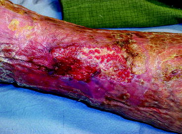
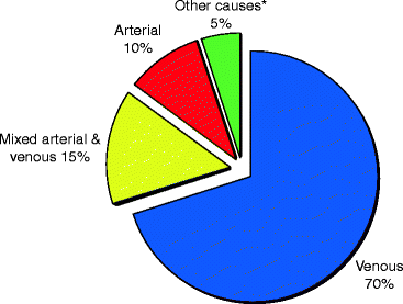
The vast majority of chronic leg ulceration will have a vascular aetiology, with chronic venous insufficiency (Fig. ).
Fig. 1.2
Differential diagnoses of leg ulcers. * Malignancy (12%), diabetes, rheumatoid arthritis, vasculitis, haematological disorders, trauma
Assessment
It is essential to differentiate between arterial and venous ulcers not only to get a precise diagnosis but also because the mainstay of treatment of venous ulcers compression therapy [].
Developments in ultrasound imaging over the past quarter of a century have had a dramatic impact on non-invasive investigations of the bodys vasculature. Duplex ultrasound imaging is useful in the assessment of venous reflux in patients with chronic leg ulceration [].
In some patients, it will be important to establish the relative importance of superficial and deep venous reflux in order to predict the effect of treating any superficial reflux. Objective measurement of the haemodynamics of venous return may be made using ambulatory venous pressure measurement (AVP). This invasive technique is done using a small needle into one of the veins on dorsum of foot and connecting the needle through a transducer to a blood pressure measurement machine. Digital photoplethysmography (PPG) is a sensitive, non-invasive test that can be performed by non-medical staff using cheap, portable equipment and provides functional information equivalent to the gold-standard AVP measurement []. The importance accurate assessments for a reliable diagnosis to ensure selection of the most effective treatment pathway for the patient and cannot be overstated.
Doppler Ultrasound Examination
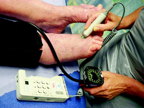
The use of handheld Doppler ultrasound as a diagnostic tool is now mandatory when assessing patients with leg ulcers. Objective measurement of the ABPI using a hand held Doppler probe is important and easily repeated [].
Fig. 1.3
Hand-held Doppler ultrasound assessment of arteries
The Doppler phenomenon states that the frequency of an incident light or sound wave is altered when it is incident on a moving object (in this case a red blood cell or pulsating blood vessels). The change in Doppler frequency due to the relative motion between the observer and the object is known as the Doppler shift []. The Doppler shift ( f D) can be calculated with the following formula:
where v is the speed of the moving target, f t is the frequency of the emitted pulse (i.e. the frequency of the transducer), is the angle between the direction of emitted sound wave and the direction of the moving target, and c is the average speed of sound within the tissue [].
In moving blood, it is the red blood cells that reflect the sound waves that result in the Doppler shifted echo. This reflection, or backscattering as it is often referred to in the texts [].
In blood vessels, the Doppler shift is dependent on the speed of blood flow, the angle between the transducer and the vessel, and the operating frequency of the Doppler transducer []. Thus, interpretation of the audible response can be susceptible to user error if the angle between the transducer beam and the moving target (i.e. blood flow) is too great. When the probe is directed precisely along the direction of flow, the Doppler shift is maximum since the angle is zero and cosine is 1. If the transducer is held at 90 to the blood vessel there will be no component of velocity along the beam (cosine is zero) and therefore no detectable Doppler shift.
Validity and Reliability of Doppler
ABPI may be derived using a handheld Doppler probe accurately. This is essentially a comparison between the systolic blood pressure in the arm and in the lower leg and is illustrated in the following formula:
In a normal subject the pressure at the ankle is often slightly higher than at the brachial fossa as there is reflection of the pulse pressure from the vascular bed of the feet. The shape of the arterial pressure pulse wave changes from the ascending aorta to the periphery; the systolic pressure increases while diastolic pressure falls at peripheral sites. A range of 0.851.25 may be considered to be consistent with the presence of normal peripheral arterial supply (see Table ).

