• Complain

Comstock Christopher E. - Breast MRI Teaching Atlas

Here you can read online Comstock Christopher E. - Breast MRI Teaching Atlas full text of the book (entire story) in english for free. Download pdf and epub, get meaning, cover and reviews about this ebook. City: New York;NY, year: 2017, publisher: Springer New York, genre: Romance novel. Description of the work, (preface) as well as reviews are available. Best literature library LitArk.com created for fans of good reading and offers a wide selection of genres:

Romance novel Science fiction Adventure Detective Science History Home and family Prose Art Politics Computer Non-fiction Religion Business Children Humor

Choose a favorite category and find really read worthwhile books. Enjoy immersion in the world of imagination, feel the emotions of the characters or learn something new for yourself, make an fascinating discovery.

Comstock Christopher E. Breast MRI Teaching Atlas

Breast MRI Teaching Atlas: summary, description and annotation

We offer to read an annotation, description, summary or preface (depends on what the author of the book "Breast MRI Teaching Atlas" wrote himself). If you haven't found the necessary information about the book — write in the comments, we will try to find it.

This atlas serves as a basic introduction to breast MRI. Organized by case, it emphasizes pertinent breast MRI findings and common indications for breast MRI. Topics include breast MRI basics, benign findings, breast malignancy, high-risk conditions, interesting cases, and breast implants. Brief teaching points accompany each case and highlight the importance of the findings. Breast MRI Teaching Atlas is an ideal resource for diagnostic radiologists, residents, fellows, and clinicians involved in the care of breast cancer patients, including surgeons, oncologists, and obstetricians/gynecologists. -- Prov de leditor.

Comstock Christopher E.: author's other books


Who wrote Breast MRI Teaching Atlas? Find out the surname, the name of the author of the book and a list of all author's works by series.

Breast MRI Teaching Atlas — read online for free the complete book (whole text) full work

Below is the text of the book, divided by pages. System saving the place of the last page read, allows you to conveniently read the book "Breast MRI Teaching Atlas" online for free, without having to search again every time where you left off. Put a bookmark, and you can go to the page where you finished reading at any time.

Light

Font size:

Reset

Interval:

Bookmark:

Make
Springer New York 2017
Richard Ha , Christopher E. Comstock and Elizabeth A. Morris (eds.) Breast MRI Teaching Atlas 10.1007/978-1-4939-6409-3_1
1. Breast MRI Basics
Victoria Mango 1
(1)
Breast Imaging Service, Memorial Sloan-Kettering Cancer Center, Assistant Professor of Radiology, Weill Cornell Medical College, New York, NY, USA
(2)
Breast Imaging Section, Associate Professor of Radiology, University of Washington School of Medicine, Seattle, Washington, USA
(3)
Division of Breast Imaging, Associate Professor of Radiology, Columbia University Medical Center, New York, NY, USA
(4)
Division of Breast Imaging, Assistant Professor of Radiology, Columbia University Medical Center, New York, NY, USA
(5)
Division of Breast Imaging, Assistant Professor of Radiology, Columbia University Medical Center, New York, NY, USA
Victoria Mango Director of Breast and Body Imaging Fellowship
Email:
Habib Rahbar
Email:
Ralph Wynn Section Head
Email:
Lauren Friedlander Director of Breast and Body Imaging Fellowship
Email:
Richard Ha Director of Education and Research (Corresponding author)
Email:
Keywords
Fibroglandular tissue Background parenchymal enhancement Maximum intensity projection Kinetics
1.1 History
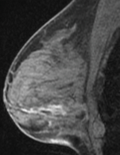
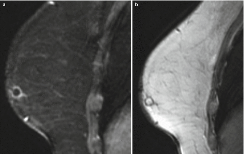
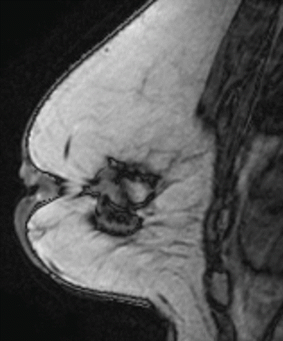
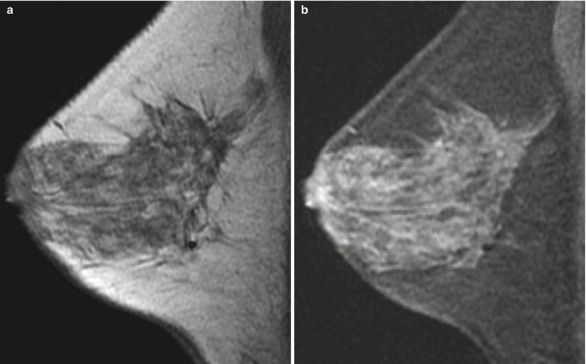
Four patients with T1-weighted sequence findings (Figs. ).
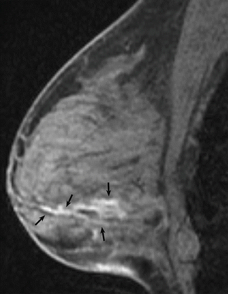
Fig 11 Sagittal T1 fat-saturated pre-contrast image of the right breast - photo 1
Fig. 1.1
Sagittal T1 fat-saturated pre-contrast image of the right breast
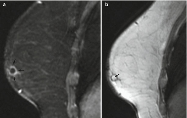
Fig 12 Sagittal T1 fat-saturated post-contrast image of the right breast a - photo 2
Fig. 1.2
Sagittal T1 fat-saturated post-contrast image of the right breast ( a ) and corresponding T1 nonfat-saturated image ( b )
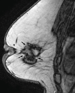
Fig 13 Sagittal T1 nonfat-saturated image of the left breast Fig 14 - photo 3
Fig. 1.3
Sagittal T1 nonfat-saturated image of the left breast
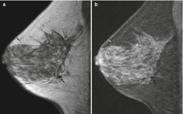
Fig 14 Sagittal T1 nonfat-saturated image of the right breast a and - photo 4
Fig. 1.4
Sagittal T1 nonfat-saturated image of the right breast ( a ) and corresponding T1 fat-saturated sequences ( b )
1.2 T1-Weighted Sequence
Teaching Points
Breast MRI should be performed on systems with at least 1.5 Tesla magnet strength with a dedicated breast coil. Usually, a pre-contrast T1-weighted gradient-echo sequence without fat saturation is the first sequence performed after the scout images. A T1-weighted nonfat-saturated sequence is obtained bilaterally, including axillae and chest wall, to distinguish fat from water-based tissues. The pre-contrast T1-weighted sequence is typically used to assess the amount of fibroglandular tissue and can be particularly helpful in documenting the presence of high-signal hemorrhagic or proteinaceous fluid-filled, dilated ducts (Fig. ).
Image Findings
Fig 15 Hemorrhagic or proteinaceous debris in ducts Sagittal T1 - photo 5
Fig. 1.5
Hemorrhagic or proteinaceous debris in ducts. Sagittal T1 fat-saturated pre-contrast image of the right breast demonstrates several linear hyperintensities ( arrows ) in a ductal distribution in the lower breast, compatible with hemorrhagic or proteinaceous debris
Fig 16 Fat necrosis a Sagittal T1 fat-saturated post-contrast image of - photo 6
Fig. 1.6
Fat necrosis. ( a ) Sagittal T1 fat-saturated post-contrast image of the right breast demonstrates indeterminate rim enhancement ( arrows ). ( b ) The T1 nonfat-saturated image shows central fat ( arrow ), compatible with benign fat necrosis
Fig 17 Architectural distortion In a patient with prior lumpectomy the - photo 7
Fig. 1.7
Architectural distortion. In a patient with prior lumpectomy, the sagittal T1 nonfat-saturated image of the left breast illustrates architectural distortion in the central breast ( arrows ) and associated nipple retraction ( arrowhead )
Fig 18 Susceptibility artifact from biopsy clip The susceptibility artifact - photo 8
Fig. 1.8
Susceptibility artifact from biopsy clip. The susceptibility artifact from a biopsy clip ( arrow ) is more prominent on the T1 nonfat-saturated image of the right breast ( a ) than on the T1 fat-saturated sequences ( b )
1.3 History
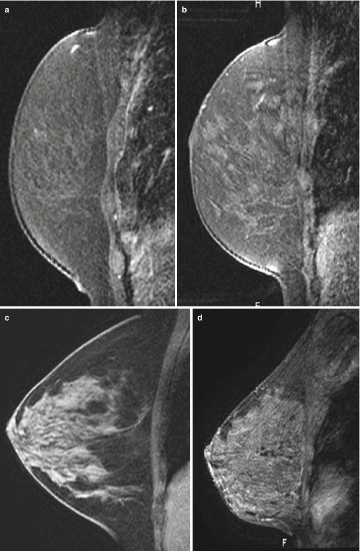
Four patients illustrating the range of the amount of fibroglandular tissue on breast MRI (Figs. ).
Fig 19 110 111 and 112 Selected sagittal T1 post-contrast image a - photo 9
Fig. 1.9, 1.10, 1.11, and 1.12
Selected sagittal T1 post-contrast image ( a ) right breast demonstrates an almost entirely fatty breast ( b ) right breast shows scattered fibroglandular tissue. Selected sagittal T1 pre-contrast image ( c ) right breast demonstrates heterogeneous fibroglandular tissue. Selected sagittal T1 post-contrast image ( d ) left breast demonstrates extreme fibroglandular tissue
1.4 Fibroglandular Tissue
Teaching Points
At mammography, breast density is represented by the amount of fibroglandular tissue (FGT) in contrast to fat measured in two-dimensional views that may not reflect an accurate assessment. Breast MRI provides strong soft-tissue contrast between FGT and fat, and a more accurate three-dimensional coverage of the entire breast. Evaluation of the amount of FGT is usually done on the T1-weighted sequences with and without fat suppression. According to the American College of Radiology (ACR) guidelines, the amount of FGT should be described in the Breast MRI report using the BI-RADS four assessment categories of breast composition, which are defined by the visually estimated content of FGT within the breasts.
The amount of fibroglandular tissue (breast density) has been established as an independent risk factor associated with the development of breast cancer, which is three to five times higher in women with mammographically dense breasts than in women with predominantly fatty breasts.
1.5 History
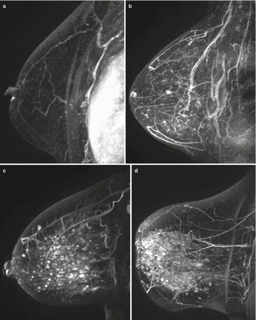
Four patients illustrating the range of background parenchymal enhancement seen on breast MRI (Figs. ).
Fig 113114 115 and 116 Sagittal T1 post-contrast 3D maximum intensity - photo 10
Fig. 1.13,1.14, 1.15, and 1.16
Sagittal T1 post-contrast 3D maximum intensity projection (MIP) image ( a ) left breast demonstrates minimal background parenchymal enhancement ( b ) right breast demonstrates mild background parenchymal enhancement ( c ) right breast demonstrates moderate background parenchymal enhancement ( d ) right breast demonstrates marked background parenchymal enhancement
Next page
Light

Font size:

Reset

Interval:

Bookmark:

Make

Similar books «Breast MRI Teaching Atlas»

Look at similar books to Breast MRI Teaching Atlas. We have selected literature similar in name and meaning in the hope of providing readers with more options to find new, interesting, not yet read works.


Reviews about «Breast MRI Teaching Atlas»

Discussion, reviews of the book Breast MRI Teaching Atlas and just readers' own opinions. Leave your comments, write what you think about the work, its meaning or the main characters. Specify what exactly you liked and what you didn't like, and why you think so.