• Complain

Sonnenschein Martin - Atlas of Breast Tomosynthesis Imaging Findings and Image-Guided Interventions

Here you can read online Sonnenschein Martin - Atlas of Breast Tomosynthesis Imaging Findings and Image-Guided Interventions full text of the book (entire story) in english for free. Download pdf and epub, get meaning, cover and reviews about this ebook. City: Cham, year: 2017;2018, publisher: Springer International Publishing, genre: Home and family. Description of the work, (preface) as well as reviews are available. Best literature library LitArk.com created for fans of good reading and offers a wide selection of genres:

Romance novel Science fiction Adventure Detective Science History Home and family Prose Art Politics Computer Non-fiction Religion Business Children Humor

Choose a favorite category and find really read worthwhile books. Enjoy immersion in the world of imagination, feel the emotions of the characters or learn something new for yourself, make an fascinating discovery.

Sonnenschein Martin Atlas of Breast Tomosynthesis Imaging Findings and Image-Guided Interventions

Atlas of Breast Tomosynthesis Imaging Findings and Image-Guided Interventions: summary, description and annotation

We offer to read an annotation, description, summary or preface (depends on what the author of the book "Atlas of Breast Tomosynthesis Imaging Findings and Image-Guided Interventions" wrote himself). If you haven't found the necessary information about the book — write in the comments, we will try to find it.

Sonnenschein Martin: author's other books


Who wrote Atlas of Breast Tomosynthesis Imaging Findings and Image-Guided Interventions? Find out the surname, the name of the author of the book and a list of all author's works by series.

Atlas of Breast Tomosynthesis Imaging Findings and Image-Guided Interventions — read online for free the complete book (whole text) full work

Below is the text of the book, divided by pages. System saving the place of the last page read, allows you to conveniently read the book "Atlas of Breast Tomosynthesis Imaging Findings and Image-Guided Interventions" online for free, without having to search again every time where you left off. Put a bookmark, and you can go to the page where you finished reading at any time.

Light

Font size:

Reset

Interval:

Bookmark:

Make
Springer International Publishing Switzerland 2017
Martin Sonnenschein and Christian Waldherr Atlas of Breast Tomosynthesis 10.1007/978-3-319-21566-2_1
1. Introduction to Tomosynthesis
Martin Sonnenschein 1 and Christian Waldherr 1
(1)
Bern, Switzerland
1.1 Motivation for Tomosynthesis
Mammography is X-ray imaging of the breast. The technique works because different components of the breast absorb X-rays in different amounts, generating an image in which fat, fibrous, and glandular tissue; soft tissue lesions; and calcifications can be identified. Fibroglandular tissue and soft tissue lesions have almost identical X-ray absorption properties, which can make identifying small cancers challenging when using mammography. Unless a lesion is located in a region that is surrounded by fat, its visibility is reduced because of the shadows of fibroglandular structures overlying the cancer. These interfering structures are sometimes referred to as structure noise or anatomical noise. In denser breasts, there are more structures that interfere with seeing a cancer, and the clinical performance of mammography decreases with increasing breast density (Pisano et al. ). The sensitivity, or ability to detect cancer, is lower in extremely dense breasts compared with breasts consisting mainly of fat.
A limitation of mammography is that it is a two-dimensional (2D) imaging modality. Fibroglandular tissues can hide small cancers, reducing sensitivity, and the noise caused by the fibroglandular tissues can either mimic a cancer or render an accurate benign diagnosis difficult, reducing specificity. One way to improve the sensitivity and specificity of X-ray imaging is to perform three-dimensional (3D) imaging, which can reduce the confounding effects of fibroglandular tissues on cancer detection, especially in denser breasts (Niklason et al. ). This technique is called breast tomosynthesis and is also known as digital breast tomosynthesis (DBT), 3D mammography, and tomosynthesis.
1.2 The Physics of Breast Tomosynthesis Imaging
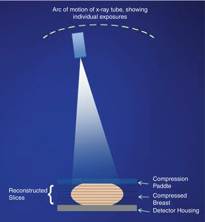
Figure presents the basics of a breast tomosynthesis system. The breast is compressed, as with conventional 2D mammography, between a compression paddle and the detector housing. While the breast is kept stationary, the X-ray tube is moved, usually in an arcuate motion, and a set of low-dose images known as projections is collected. These are acquired at different angular locations of the tube. The total angular range covered by the X-ray tube is called the scan angle.
Fig 11 Schematic showing of the basic operation of a tomosynthesis system - photo 1
Fig. 1.1
Schematic showing of the basic operation of a tomosynthesis system
After the scan is completed, the projections are used to calculate a 3D image consisting of high-resolution tissue slices whose planes are parallel to the breast support plates. This procedure is known as reconstruction and is similar to the methods used to reconstruct images from a conventional computed tomography (CT) scanner. The number of reconstructed slices depends on the thickness of the compressed breast and the desired separation between slices, typically about 1 mm. After reconstruction, the slices are transmitted to a diagnostic workstation for display and review by a radiologist.
1.2.1 Acquisition and Reconstruction Methods
The way in which tomosynthesis acquisition is performed varies from manufacturer to manufacturer. For commercially available systems or those that have been evaluated in prototype versions, scan angles range from 11 to 50, reconstructed slice thicknesses range from 0.5 to 1.0 mm, and scan times range from less than 4 to 25 s. A variety of reconstruction algorithms have been evaluated, including filtered back projection and iterative reconstruction methods. For the purposes of this book, with its clinical emphasis, the system variations mentioned here may not be relevant. These systems all generate 3D images that can be viewed in thin, cross-sectional slices through the breast.
1.2.2 Characteristics of the Reconstructed Image
Tomosynthesis is a form of limited-angle tomography. The term limited angle means that, unlike CT imaging, where the X-ray source and detector rotate through 360, the smaller scan angles in breast tomosynthesis result in image characteristics that are fundamentally different from a CT image. In a CT image, the voxels, or three-dimensional pixel, can be isotropic. In other words, the x , y , and z dimensions of the voxel are similar in size. In a breast tomosynthesis system, the x and y dimensions, or resolution, are often finer than in a CT image, and the third dimension, z, has a resolution that is poorer than the x and y resolution. Typical values for a breast tomosynthesis system exhibit an x and y resolution of about 0.1 mm and a z resolution of about 35 mm. Confusingly, the z resolution is not the same as the slice thickness, which is typically 1 mm. When it is said that the z resolution is 3 mm, for example, it can be observed that an object will be visible in several slices above and below its actual location in space. If reconstruction results in a thinner slice, say 0.5 mm, then the object will be visible in twice as many slices. Making the reconstructed slice thinner does not improve the z resolution of the system.
The nonisotropic nature of the voxels defines a natural coordinate system, which is where slices are parallel to the breast platform. A craniocaudal (CC) image cannot be reformatted into a mediolateral oblique (MLO) image, or vice versa, because the resolution in the z direction is poorer than the resolution in the x , y plane. This differs from CT imaging, where an image can easily be reformatted into a transverse, sagittal, coronal, or any arbitrary orientation.
There are important clinical ramifications to the nonisotropic nature of the image. Tumors may appear more conspicuous in one projection than another, and multiple studies have shown that cancers are often better visualized in either the CC or MLO tomosynthesis image; therefore, acquiring both CC and MLO 3D images may maximize cancer detection (Rafferty et al. ). This is analogous to the standard practice in 2D mammography.
1.3 Synthesized 2D Images
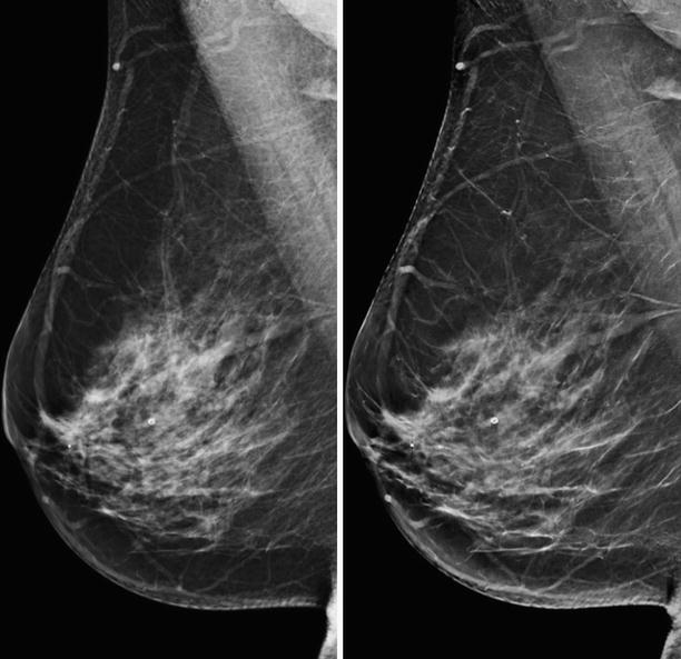
The first screening trials employing breast tomosynthesis used a protocol in which tomosynthesis was added as an adjunct to 2D imaging, that is, breasts were imaged with both 2D and 3D images, the so-called combo mode: 2D-CC and 2D-MLO and 3D-CC and 3D-MLO (Skaane et al. ). Thus, 2D images, the standard of care, were available for comparison with previous 2D images, and 3D images were available to provide additional information. This approach demonstrated good clinical performance, the only downside being the additional radiation dose delivered because both two-view 2D and two-view 3D images were acquired. Manufacturers have sought ways to reduce the dose, one approach being to eliminate the 2D images by generating them directly from the 3D images. This returns the radiation dose to a level comparable to standard 2D mammography.
Fig 12 Left image is a 2D mammogram and the right image is a 2D image - photo 2
Fig. 1.2
Left image is a 2D mammogram, and the right image is a 2D image synthesized from the 3D image set
The algorithms used to create the synthesized 2D image create a 2D-like image that is similar, but not identical, to a true 2D image. It is possible to enhance the visibility of objects such as microcalcifications and architectural distortions so that they are more conspicuous than they are in a conventional 2D image.
Next page
Light

Font size:

Reset

Interval:

Bookmark:

Make

Similar books «Atlas of Breast Tomosynthesis Imaging Findings and Image-Guided Interventions»

Look at similar books to Atlas of Breast Tomosynthesis Imaging Findings and Image-Guided Interventions. We have selected literature similar in name and meaning in the hope of providing readers with more options to find new, interesting, not yet read works.


Reviews about «Atlas of Breast Tomosynthesis Imaging Findings and Image-Guided Interventions»

Discussion, reviews of the book Atlas of Breast Tomosynthesis Imaging Findings and Image-Guided Interventions and just readers' own opinions. Leave your comments, write what you think about the work, its meaning or the main characters. Specify what exactly you liked and what you didn't like, and why you think so.