• Complain

Grabherr Silke - Atlas of Postmortem Angiography

Here you can read online Grabherr Silke - Atlas of Postmortem Angiography full text of the book (entire story) in english for free. Download pdf and epub, get meaning, cover and reviews about this ebook. City: Cham, year: 2016;2018, publisher: Springer International Publishing, genre: Romance novel. Description of the work, (preface) as well as reviews are available. Best literature library LitArk.com created for fans of good reading and offers a wide selection of genres:

Romance novel Science fiction Adventure Detective Science History Home and family Prose Art Politics Computer Non-fiction Religion Business Children Humor

Choose a favorite category and find really read worthwhile books. Enjoy immersion in the world of imagination, feel the emotions of the characters or learn something new for yourself, make an fascinating discovery.

Grabherr Silke Atlas of Postmortem Angiography

Atlas of Postmortem Angiography: summary, description and annotation

We offer to read an annotation, description, summary or preface (depends on what the author of the book "Atlas of Postmortem Angiography" wrote himself). If you haven't found the necessary information about the book — write in the comments, we will try to find it.

Grabherr Silke: author's other books


Who wrote Atlas of Postmortem Angiography? Find out the surname, the name of the author of the book and a list of all author's works by series.

Atlas of Postmortem Angiography — read online for free the complete book (whole text) full work

Below is the text of the book, divided by pages. System saving the place of the last page read, allows you to conveniently read the book "Atlas of Postmortem Angiography" online for free, without having to search again every time where you left off. Put a bookmark, and you can go to the page where you finished reading at any time.

Light

Font size:

Reset

Interval:

Bookmark:

Make
Part I
Introduction
Springer International Publishing Switzerland 2016
Silke Grabherr , Jochen M. Grimm and Axel Heinemann (eds.) Atlas of Postmortem Angiography 10.1007/978-3-319-28537-5_1
1. Postmortem Imaging: Development and Historical Review
Adrian M. K. Thomas 1
(1)
Faculty of Health and Sciences, Canterbury Christ Church University, Canterbury, Kent, UK
Adrian M. K. Thomas
Email:
Keywords
Death Memento mori Postmortem angiography
With the development of Virtopsy in the 1990s, it would be reasonable to imagine that postmortem imaging is a recent development. In fact, postmortem imaging has been part of radiology since the days of the pioneers. The reality is that postmortem imaging was practiced right at the start of imaging. As new radiologic techniques have developed they have been applied to postmortem studies, and postmortem studies have assisted in the development of radiologic techniques.
1.1 The Discovery of X-rays
1.1.1 Wilhelm Rntgen and the Discovery of X-rays
During the nineteenth century there was considerable scientific interest in passing electrical currents across fluids, and this was pioneered by Michael Faraday at the Royal Institution in London. These fluids could be liquids or gases, and as the century progressed there was an increasing ability to produce a progressively higher vacuum in a glass bulb.
In 1895 Wilhelm Conrad Rntgen, who was a 50-year-old physics professor at the University of Wrzburg in Germany [], made his classic experiment on the afternoon of 8 November. Rntgen was using a covered Hittorf-Crookes tube (an evacuated glass bulb), and he observed the fluorescence of a screen at a distance. This was the origin of what was to be called fluoroscopy.
The famous radiograph that Rntgen made of his wife Berthas hand was taken on 22 December 1895, and Rntgen delivered his preliminary report On a New Kind of Rays ( ber eine neue Art von Strahlen ) to the Physical-Medical Society of Wrzburg on December 28th 1895 for publication in their proceedings []. Although at the time this discovery could be seen as entirely unexpected, with hindsight we can discern a certain inevitability about it.
The news reached the scientific community in the first week of 1896 by two main routes []. First, Wilhelm Rntgen had sent reprints of his paper and samples of his radiographs to his physicist colleagues throughout the world. Second, most of the public heard of the discovery through the daily newspapers from foreign correspondents who had picked up the news of the discovery from the Vienna paper Neue Freie Presse . The papers editors son was a young physicist colleague of Franz Exner, another friend of Rntgen who had received a reprint of the first communication. Although the short newspaper reports of January 1896 were factual and contained limited associated commentary, their significance was not lost on those who were familiar with recent advances in experimental science. No previous scientific discovery had such a dramatic reception as that which greeted Rntgens work. The popular press, the world of science, and the medical profession reacted with a mixture of amazement, incredulity, and applause.
Although the first radiograph that Rntgen obtained was that of the hand of his wife Bertha, he soon realized that radiography could be used in other areas. Other items he radiographed included a coil of wire, a box of weights, and the door of his laboratory. These observations were to have great significance for forensic radiology and for nondestructive testing. In the summer of 1896, Rntgen took a radiograph of his shotgun, which he sent to Franz Exner in Vienna.
Rntgen was honored with the first Nobel Prize in physics awarded in 1901.
1.1.2 Popular Reception of the Discovery
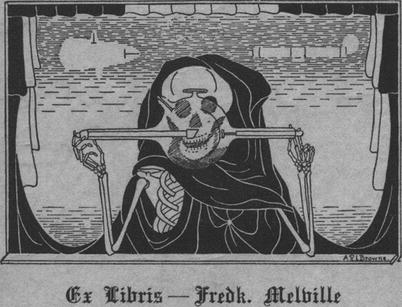
It is now quite difficult for us to understand the complete astonishment with which the discovery of x-rays was greeted by the general public. However, there was something decidedly macabre about being able to visualize the bones of the body during life, and radiography became a memento mori, a reminder that we are mortal and that we all one day will die, as shown in the bookplate of the pioneer Frederick Melville (Fig. ). In the modern world we are now so used to seeing medical images in contemporary media that the sense of fear is considerably less marked. Prior to medical imaging the inside of the body was seen only in the graveyard, battlefield, or the operating theater.
Fig 11 The bookplate of Frederick Melville The x-ray tube reveals death the - photo 1
Fig. 1.1
The bookplate of Frederick Melville. The x-ray tube reveals death, the grim reaper (Public domain.)
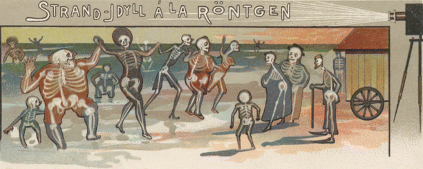
Fig 12 Strand-Idyll la Rntgen The x-rays reveal the dancing skeletons in - photo 2
Fig. 1.2
Strand-Idyll la Rntgen. The x-rays reveal the dancing skeletons in this otherwise charming beach scene. Notice the varying sizes of the skeletons, depending on the human morphology! (Public domain.)
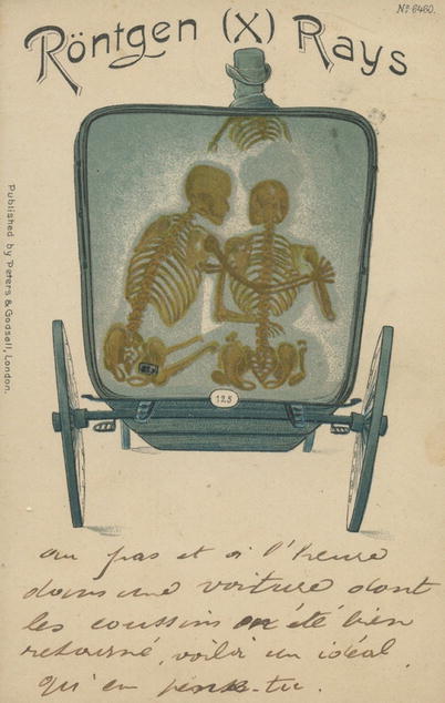
Fig 13 Rntgen X rays The x-rays show the couple in the taxi as skeletons - photo 3
Fig. 1.3
Rntgen (X) rays. The x-rays show the couple in the taxi as skeletons in this postcard (Public domain.)
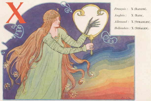
Fig 14 X rayons The beautiful girl sees her deaths hand on the fluorescent - photo 4
Fig. 1.4
X (rayons). The beautiful girl sees her deaths hand on the fluorescent screen (Public domain.)
1.2 Pioneer Radiology
Although the first image obtained was of a living hand, it soon became obvious that the new rays could be useful in looking at nonliving tissue. The x-rays were rapidly used throughout the world for two reasons. First Rntgens discovery had been rapidly communicated both by Rntgen himself and by newspapers, magazines, and scientific journals. It should also be remembered that the apparatus needed was already present in almost all physics departments, and the Hittorf-Crookes tube could be easily obtained.
1.2.1 Role of Postmortem Imaging
  • Simple curiosity.
  • Development of radiologic techniques.
  • Use as a radiologic phantom/test object.
  • Increased anatomic knowledge.
  • Use in forensic medicine.
  • Anthropological research.
  • Archeological research.
1.2.2 Charles Thurstan Holland
There were many pioneers in the early days of radiology. The pioneer use of radiology is particularly well illustrated in the work of Charles Thurstan Holland.
In the paper X-Rays in 1896 by Charles Thurstan Holland [], he described his radiologic work in 1896. He was a pioneer radiologist in Liverpool in the United Kingdom and was the President of the First International Congress of Radiology held in London in 1925. The work that Holland did in that first year and with his primitive apparatus is quite remarkable. His interest in radiology was attributable to the encouragement of the orthopedic surgeon Sir Robert Jones.
In practice many of the issues confronting postmortem imaging are the same as those encountered with radiography during life. The principles involved in both cases are similar. It is easy for us to think that the simple plain film images obtained in those early days must have been easy to interpret compared to the rather more complex three-dimensional images that we see today. In reality nothing could be further from the truth. Holland gives an excellent account as to exactly how difficult image interpretation was in those early days. As an example, on June 16 1896 he radiographed the forearm of a boy and demonstrated bone necrosis and bone loss. Holland said that this case opened our eyes to the big question of interpretation. Although the changes were gross, we were all wondering as to what it all meant. He radiographed a Potts fracture of the ankle and looked at the radiograph with Sir Robert Jones. They spent a long time looking at the images and again were wondering what it all meant. Whether imaging the living or the dead, the images obtained were and continue to be confusing. What is obvious to us retrospectively is not quite so obvious prospectively.
Next page
Light

Font size:

Reset

Interval:

Bookmark:

Make

Similar books «Atlas of Postmortem Angiography»

Look at similar books to Atlas of Postmortem Angiography. We have selected literature similar in name and meaning in the hope of providing readers with more options to find new, interesting, not yet read works.


Reviews about «Atlas of Postmortem Angiography»

Discussion, reviews of the book Atlas of Postmortem Angiography and just readers' own opinions. Leave your comments, write what you think about the work, its meaning or the main characters. Specify what exactly you liked and what you didn't like, and why you think so.