• Complain

Etehadtavakol Mahnaz - Application of Infrared to Biomedical Sciences

Here you can read online Etehadtavakol Mahnaz - Application of Infrared to Biomedical Sciences full text of the book (entire story) in english for free. Download pdf and epub, get meaning, cover and reviews about this ebook. City: Singapore, year: 2017, publisher: Springer Singapore, genre: Romance novel. Description of the work, (preface) as well as reviews are available. Best literature library LitArk.com created for fans of good reading and offers a wide selection of genres:

Romance novel Science fiction Adventure Detective Science History Home and family Prose Art Politics Computer Non-fiction Religion Business Children Humor

Choose a favorite category and find really read worthwhile books. Enjoy immersion in the world of imagination, feel the emotions of the characters or learn something new for yourself, make an fascinating discovery.

Etehadtavakol Mahnaz Application of Infrared to Biomedical Sciences

Application of Infrared to Biomedical Sciences: summary, description and annotation

We offer to read an annotation, description, summary or preface (depends on what the author of the book "Application of Infrared to Biomedical Sciences" wrote himself). If you haven't found the necessary information about the book — write in the comments, we will try to find it.

Etehadtavakol Mahnaz: author's other books


Who wrote Application of Infrared to Biomedical Sciences? Find out the surname, the name of the author of the book and a list of all author's works by series.

Application of Infrared to Biomedical Sciences — read online for free the complete book (whole text) full work

Below is the text of the book, divided by pages. System saving the place of the last page read, allows you to conveniently read the book "Application of Infrared to Biomedical Sciences" online for free, without having to search again every time where you left off. Put a bookmark, and you can go to the page where you finished reading at any time.

Light

Font size:

Reset

Interval:

Bookmark:

Make
Springer Nature Singapore Pte Ltd. 2017
Eddie YK Ng and Mahnaz Etehadtavakol (eds.) Application of Infrared to Biomedical Sciences Series in BioEngineering 10.1007/978-981-10-3147-2_1
Potential of Infrared Imaging in Assessing Digestive Disorders
Mahnaz Etehadtavakol 1, 2, Eddie Y. K. Ng 3 and Mohammad Hassan Emami 1, 2
(1)
Isfahan University of Medical Sciences, Isfahan, Iran
(2)
Gastrointestinal Disease Research Center, Poursina Hakim Research Institute, Isfahan, Iran
(3)
School of Mechanical and Aerospace Engineering, College of Engineering, Nanyang Technological University, 50 Nanyang Avenue, Singapore, 639798, Singapore
Eddie Y. K. Ng
Email:
Abstract
Thermography or infrared imaging is determined by detailed investigation of skin and cells temperatures . It helps clinicians to detect the regions of irregular chemical and blood vessel action in body tissue . The drive of biomedical industry with consequent rapid development in other areas of biomedical imaging has also strongly influenced the destiny of thermography in biomedical practice . During past few decades, the joint efforts of biomedical engineering and medical professionals have resulted in evolution of technological progress in infrared sensor technology, image processing, organized repository of knowledge, and their overall integration into a system. All these enabled the new tools of research and use in medical thermography. Thermography is a simple, noninvasive and reproducible test that can accurately reflect the inflammatory activities, and can be used safely and repeatedly, during biological course of inflammatory bowel disease. Objective of this study is presenting the possibility of infrared imaging in assessing digestive disorders such as irritable bowel syndrome , diverticulitis and Crohns disease .
Keywords
Thermography Digestive disorders
Introduction
Inflammatory bowel disease (IBD) influences chronic inflammation of all or part of the digestive section (Fig. ]. While during Stage III tumors have already spread to lymph nodes or Stage IV tumors have spread to more than one part of the body, improvement is difficult. However, by early detection of polyps and precancerous cells, we are able to increase chance of improvement. Therefore, a safe, noninvasive, reproducible, and standard adjunctively method that has potential for colon cancer early detection is appreciable. Common standard diagnostic tests to evaluate IBD involve clinical examination, laboratory tests, activity evidence as well as several imaging techniques.
Fig 1 Thermogram of an irritable bowel syndrome patient The first - photo 1
Fig. 1
Thermogram of an irritable bowel syndrome patient
The first documented practice of thermobiological signs happens to be the written works of Hippocrates at about 480 BC. Wet mud spread all over a patient to investigate the regions would dry first in order to achieve the hidden pathology of organs. Thereupon, by advanced studies and researches it has been confirmed that particular temperatures interrelated to organs of body were definitely indicative of healthy and unhealthy pathological mechanism []. Furthermore, a patent was filed by C. Herman in Johan Hopkins University in 2015 for 3D thermal imaging for the detection of skin lesions and other natural and abnormal conditions (US 20130116573 A1). In future work, 3D thermal imaging would be useful to visualize internal organs for promoting digestive disorders diagnosis. This paper is organized as follows: Methodology is explained in Methodology section, eighteen case reports are presented in Case Reports section, results are discussed in Results section, and Conclusion section concludes the findings.
Methodology
In this paper, presented case reports were patients with different indications of IBD and colorectal cancer. Various diagnostic tests including physical examinations, different laboratory tests, and different imaging tests were performed for each case. Different tests include the microbiology testing of stool samples, C-reactive protein, calprotectin, esophagogastroduodenoscopy, multidetector computed tomography, total colonoscopy, magnetic resonance (MR) imaging of the abdomen or MR enterocolography, upper gastrointestinal (GI) endoscopy, histopathology testing, MR enterography, and terminal ileoscopy. However, thermal imaging was performed for all the cases. Details of different tests will be explained in Case Reports section for each case separately.
2.1 Thermal Imaging Protocol Guidelines
We briefly explain some guidelines to provide thermal images to detect probably some digestive disorders []:
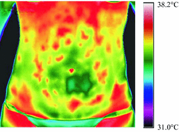
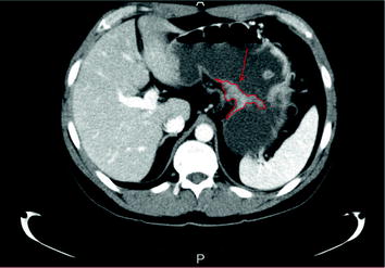
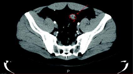
  • Imaging should be done before planning for endoscopy, to eliminate any potential influence of bowel cleansing and endoscopic mechanism.
  • Imaging can be done in two interval times: before beginning the therapy and after the patient had attained the remission, as reported by clinical, laboratory and endoscopic evaluation [].
  • The imaging itself can be progressed in this way;
    1. i.
      Patients position in front of the camera, approximately at 1 m; so that the whole abdomen was captured by the camera lens.
    2. ii.
      Patients are undressed and requested to stand in front of the camera not touching there abdomen for the sake of attaining thermal equilibrium .
    3. iii.
      The process takes about 510 min.
    4. iv.
      Consequently to attain equilibrium the patients abdomen is cooled with alcohol and then interval thermal image can be taken after equilibrium is attained again.
    5. v.
      Differences in thermal patterns and peak temperatures are captured.
    6. vi.
      Taken thermal images can be divided into four quadrants representing colonic segments: rectosigmoid, descending colon, transverse colon, and descending colon.
    7. vii.
      The same process of thermal imaging can be performed before beginning of therapy and consequently to achieve clinical remission of the disease.
    8. viii.
      Differences in observed thermal patterns and peak temperatures can be compared.
Case Reports
Case report I : In this report, a 43-year-old male patient presented to the emergency room with severe abdominal pain . Before admittance to the hospital, an esophagogastroduodenoscopy was performed that showed carcinoma on the back wall of the gastric corpus. The diagnosis of adenocarcinoma was confirmed by the pathohistological analysis. After admittance, the preoperative workup including laboratory testing and imaging methods for staging the carcinoma was performed. Laboratory testing indicated no abnormalities; however, the preformed Multidetector Computed Tomography (MDCT) indicated thickening of the gastric small curve 4 cm in diameter and a couple of affected perigastric lymph nodes. It is shown in Fig..
Fig 2 MDCT showing gastric carcinoma indicated by red part prior operation of - photo 2
Fig. 2
MDCT showing gastric carcinoma indicated by red part prior operation of a 43 years male []
Besides the observations in the stomach, the MDCT also revealed nonspecific opacification of the colon wall that could show an inflammatory process as it is demonstrated in Fig..
Fig 3 Colon diverticula and opacification of the colon wall as revealed with - photo 3
Next page
Light

Font size:

Reset

Interval:

Bookmark:

Make

Similar books «Application of Infrared to Biomedical Sciences»

Look at similar books to Application of Infrared to Biomedical Sciences. We have selected literature similar in name and meaning in the hope of providing readers with more options to find new, interesting, not yet read works.


Reviews about «Application of Infrared to Biomedical Sciences»

Discussion, reviews of the book Application of Infrared to Biomedical Sciences and just readers' own opinions. Leave your comments, write what you think about the work, its meaning or the main characters. Specify what exactly you liked and what you didn't like, and why you think so.