Joseph Sheppard - Drawing the Living Figure
Here you can read online Joseph Sheppard - Drawing the Living Figure full text of the book (entire story) in english for free. Download pdf and epub, get meaning, cover and reviews about this ebook. year: 2013, publisher: Dover Publications, genre: Romance novel. Description of the work, (preface) as well as reviews are available. Best literature library LitArk.com created for fans of good reading and offers a wide selection of genres:
Romance novel
Science fiction
Adventure
Detective
Science
History
Home and family
Prose
Art
Politics
Computer
Non-fiction
Religion
Business
Children
Humor
Choose a favorite category and find really read worthwhile books. Enjoy immersion in the world of imagination, feel the emotions of the characters or learn something new for yourself, make an fascinating discovery.
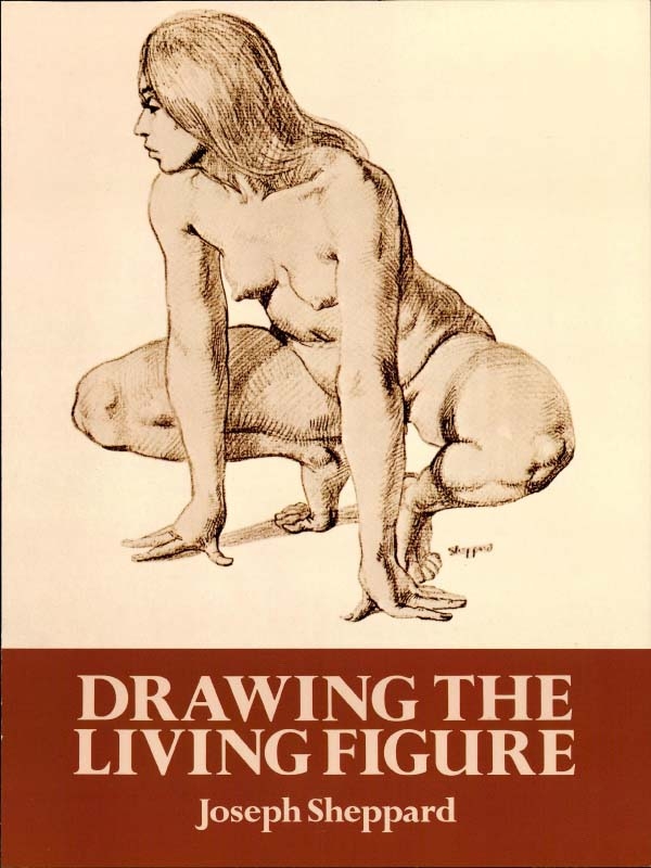
- Book:Drawing the Living Figure
- Author:
- Publisher:Dover Publications
- Genre:
- Year:2013
- Rating:5 / 5
- Favourites:Add to favourites
- Your mark:
Drawing the Living Figure: summary, description and annotation
We offer to read an annotation, description, summary or preface (depends on what the author of the book "Drawing the Living Figure" wrote himself). If you haven't found the necessary information about the book — write in the comments, we will try to find it.
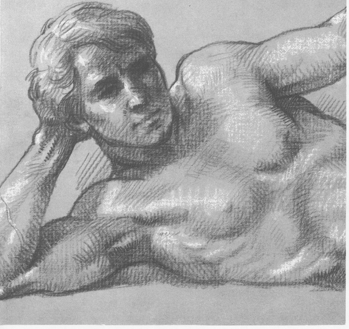
This innovative approach focuses upon the specifics of surface anatomy. 177 of Sheppards drawings show many different live models in front, back, and side views, and in various standing, sitting, kneeling, crouching, reclining, and twisting poses. Each drawing is accompanied by two diagrams, one for bones, one for muscles.
Joseph Sheppard: author's other books
Who wrote Drawing the Living Figure? Find out the surname, the name of the author of the book and a list of all author's works by series.


Drawing the Living Figure — read online for free the complete book (whole text) full work
Below is the text of the book, divided by pages. System saving the place of the last page read, allows you to conveniently read the book "Drawing the Living Figure" online for free, without having to search again every time where you left off. Put a bookmark, and you can go to the page where you finished reading at any time.
Font size:
Interval:
Bookmark:

Marsh, Reginald. Anatomy for Artists. New York: Dover Publications reprint, 1970.
Peck, Stephen Rogers. Atlas of Human Anatomy for the Artist. New York: Oxford University Press, 1951.
Richer, Paul. Artistic Anatomy. Translated and edited by Robert Beverly Hale. New York: Watson-Guptill, 1971. London: Pitman, 1973.
de C. M. Saunders, J. B. and OMalley, Charles D. The Illustrations from the Works of Andreas Vesalius. Cleveland, Ohio: The World Publishing Company, 1950.
Sheppard, Joseph. Anatomy: A Complete Guide for Artists. New York: Watson-Guptill, 1975. London: Pitman, 1975.
Sheppard, Joseph. Drawing the Female Figure. New York: Watson-Guptill, 1975. London: Pitman, 1975.
Sheppard, Joseph. Drawing the Male Figure. New York: Watson-Guptill, 1976. London: Pitman, 1976.
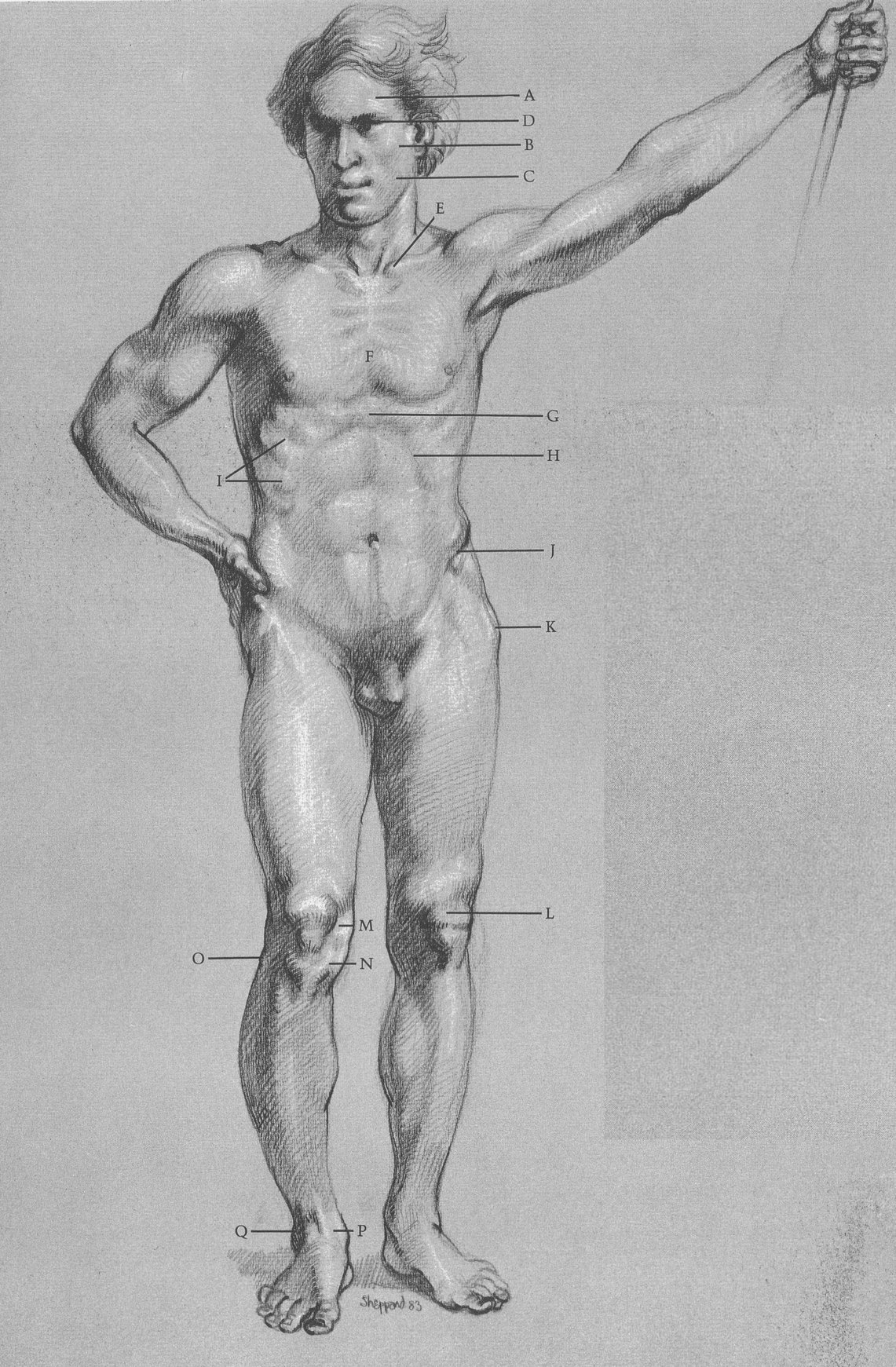
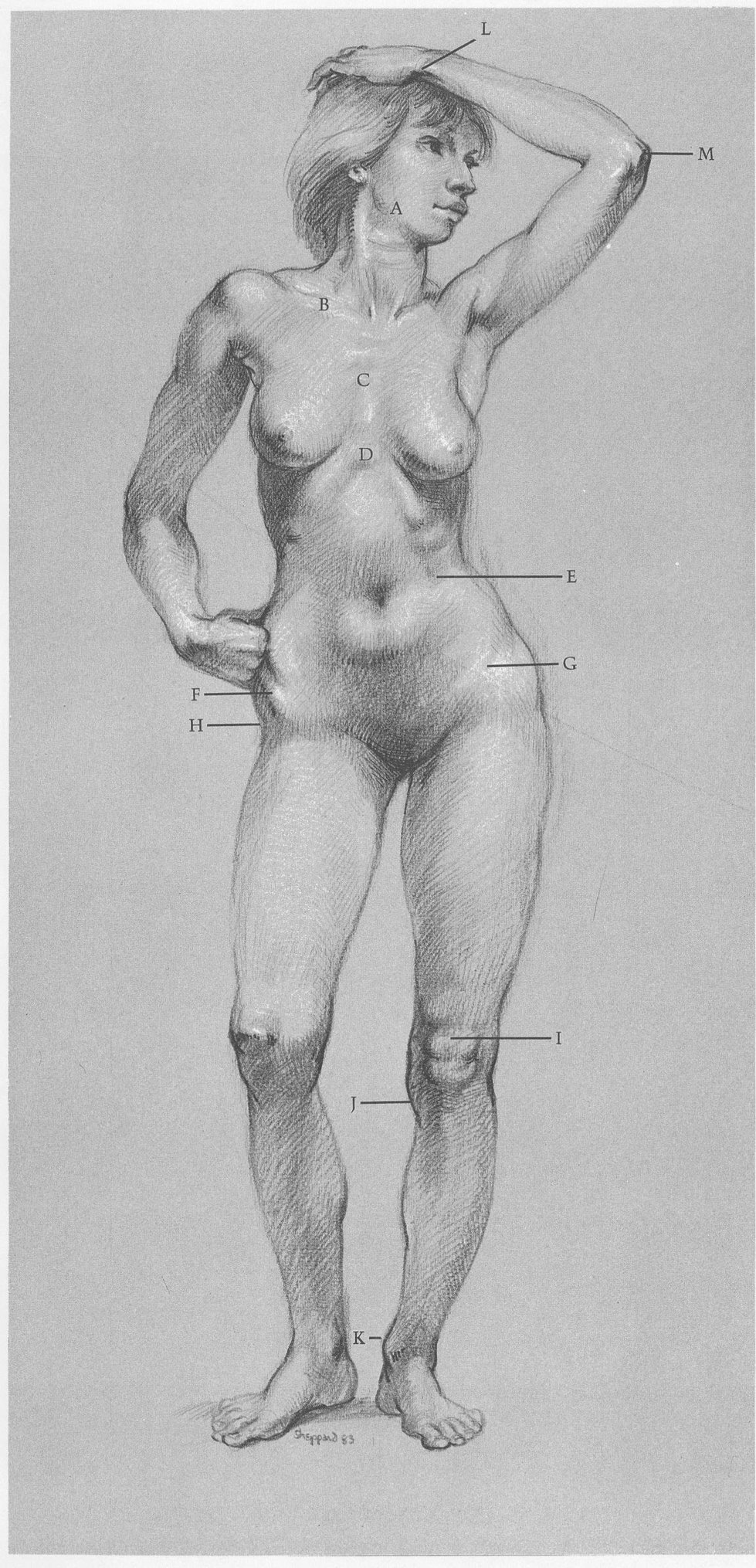
SURFACE ANATOMY
Muscles of skull are thin; bone is close to surface. Skull creates shapes of (A) forehead, (B) cheek, (C) jaw. (D) Eye is egg-shaped, sits in eye socket. (E) Clavicles start at head of (F) sternum, rising upward and back toward outside of shoulder. Note direction of first five ribs attached to sternum: first rib goes upward; second moves straight across; others point downward. (G) End of sternum protrudes. (H) Cavity of rib cage forms arch. (I) Lower ribs slant down from back to front. Pelvis holds stomach like basin. (J) Pelvic crest is prominent. (K) Hipbone is close to skin, clearly seen on male. Note oval shape of (L) kneecap. Silhouette of knee is created by (M) end of femur, (N) head of tibia. (O) Head of fibula creates bump. Ankle is formed by (P) end of tibia, (Q) end of fibula, making hinge joint for foot.
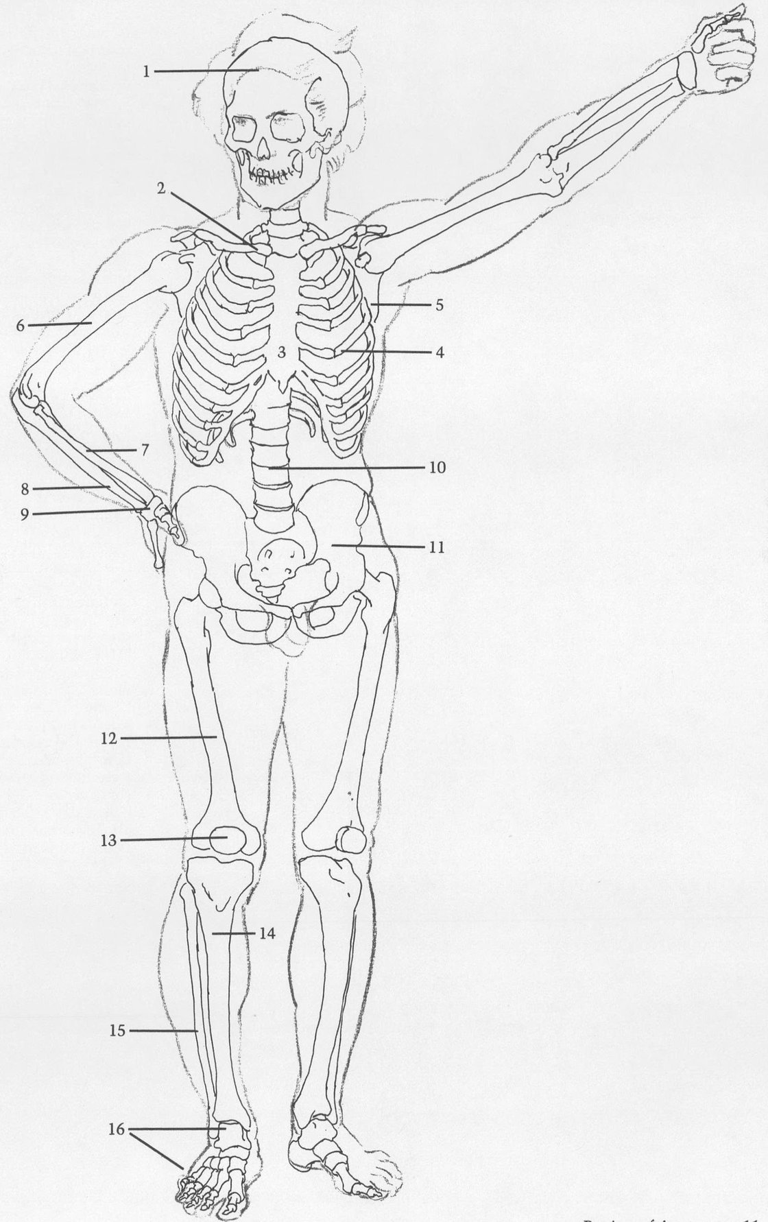
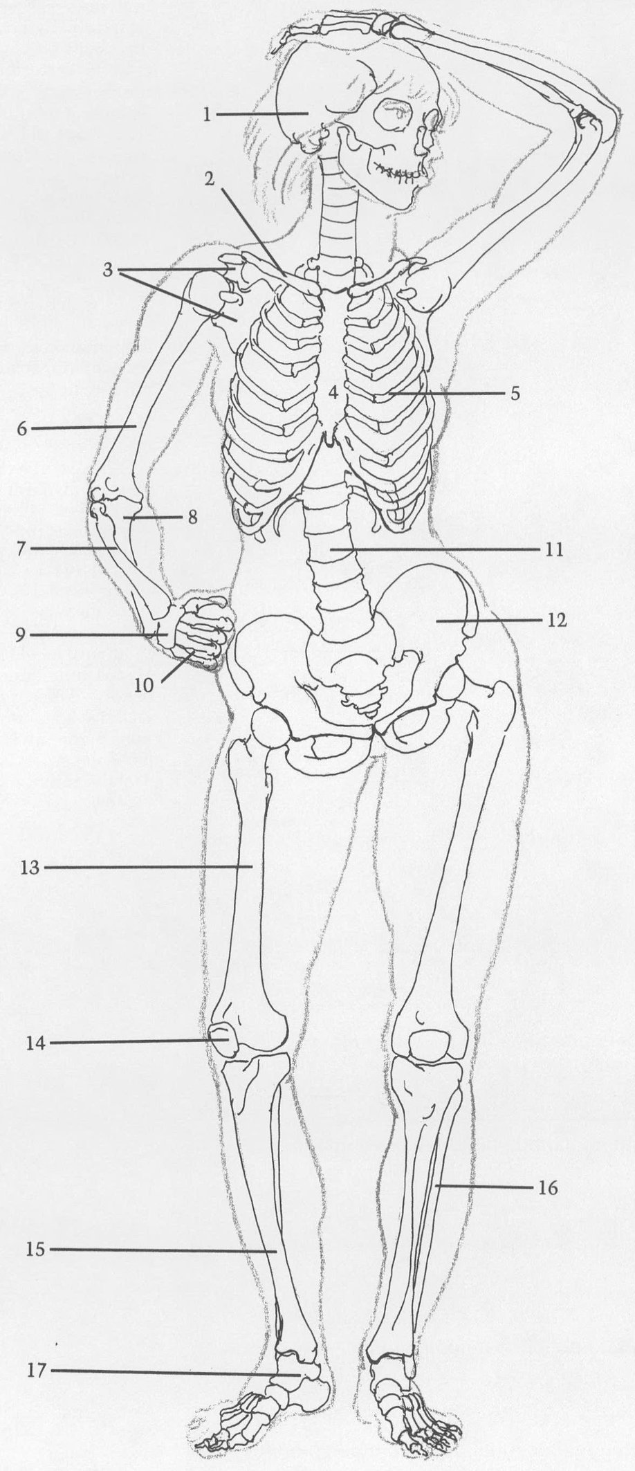
BONES
(1) Skull. (2) Clavicle. (3) Sternum. (4) Rib cage. First seven ribs attach by cartilage to sternum. Each of next three ribs attaches by cartilage to rib above. Eleventh, twelfth ribs do not attach to sternum and are called floating ribs. (5) Scapula. (6) Humerus. (7) Radius. (8) Ulna. (9) Wrist. Eight bones of wrist are treated here as one unit. (10) Spinal column is treated here as column of simple discs. (11) Pelvis. (12) Femur. (13) Kneecap. (14) Tibia. (15) Fibula attaches to rear of tibia head, intersecting tibia at ankle and descending further, thus making outside of ankle lower than inside. (16) Bones of foot.
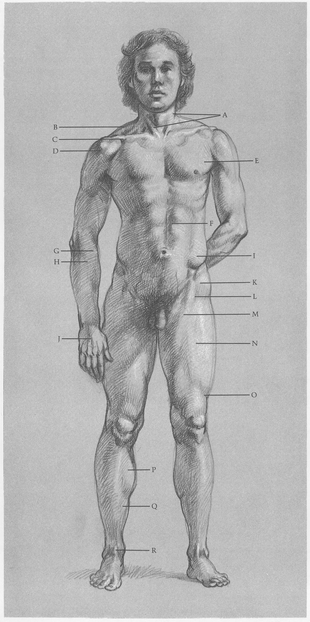
SURFACE ANATOMY
(A) Sternomastoid muscles create V shape. (B) Trapezius creates shoulder silhouette. (C) Pectoralis and deltoid meet to form cavity between them. (D) Note division of deltoid. (E) Pectoralis inserts into arm under deltoid. (F) Rectus abdominis is divided. (G) Long supinator and (H) wrist extensor both cross over from outside of elbow to thumb side of wrist. (I) External oblique inserts into top of pelvic crest. (J) Tendons of finger extensor are distinct. (K) Tensor fasciae latae angles toward outer contour. (L) See upside-down V where (M) sartorius and tensor fasciae latae overlap (N) rectus femoris. When knee is locked, (O) band of Richer pulls muscles in. (P) Gastrocnemius and (Q) soleus are calf muscles that attach in back of leg; they are seen from front. (R) Tendon of big toe extensor is prominent.
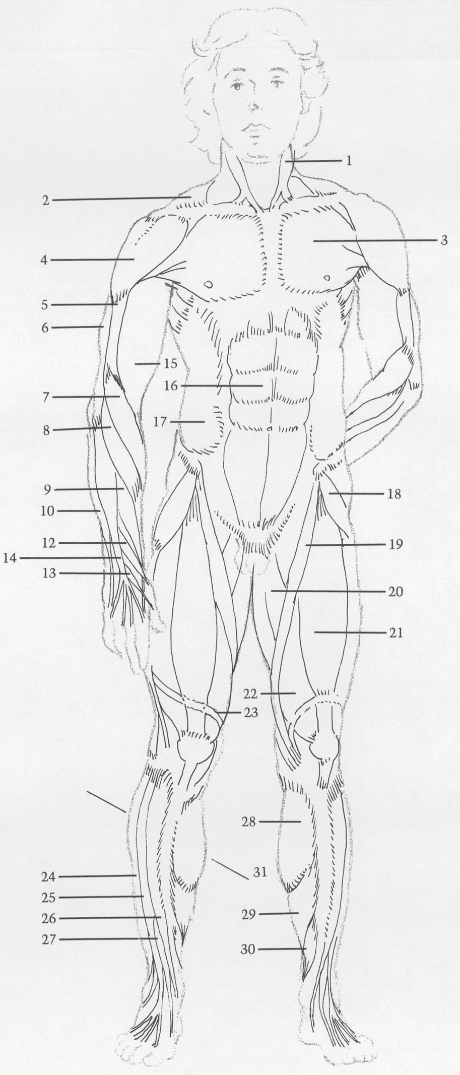
MUSCLES
(1) Stermomastoid. (2) Trapezius originates in back, inserts on top outside area of clavicle. (3) Pectoralis. (4) Deltoid. (5) Brachialis. (6) External head of triceps (one of three heads of triceps muscle). (7) Long supinator (turns forearm, palm out). (8, 9, 10) Extensors of wrist. (11) Abductor of thumb (pulls thumb toward back of hand). (12, 13) Extensors of thumb. (14) Extensor of fingers. (15) Biceps. (16) Rectus abdominis. (17) External oblique. (18) Tensor fasciae latae. (19) Sartorius. (20) Abductors. Several muscles are treated here as one unit. (21) Rectus femoris. (22) Vastus. Two parts are treated as one large muscle under rectus femoris. (23) Band of Richer changes shape of thigh when knee is locked. (Many bands that hold muscles in place are omitted). (24) Long peroneus. (25) Long extensor of toes. (26) Tibialis anterior. (27) Extensor of big toe. (28) Gastrocnemius. (29) Soleus. (30) Long flexor of toes. (31) Inside calf muscle mass is lower than outside mass.
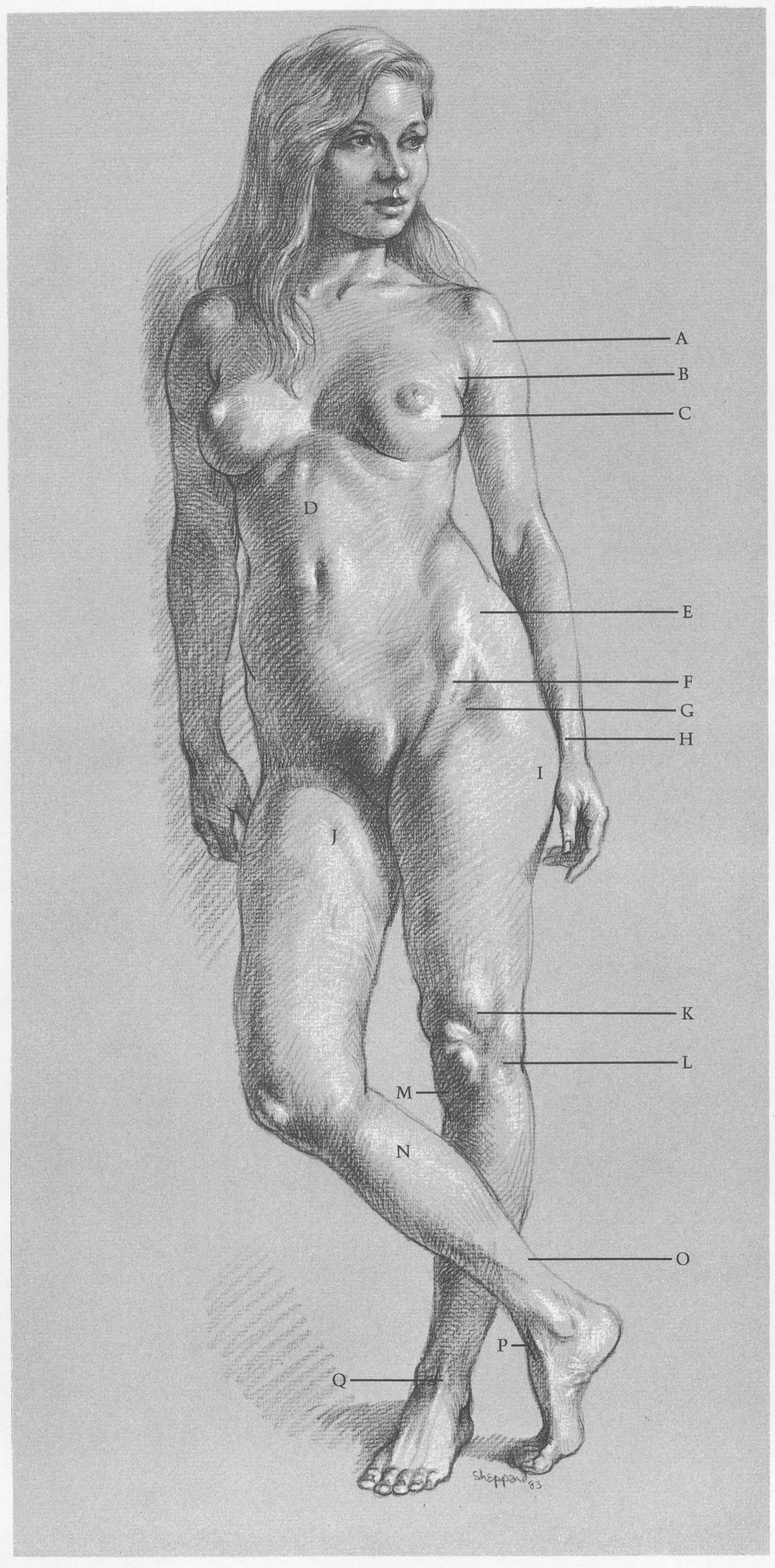
SURFACE ANATOMY
(A) Jawbone determines contour of face. (B) Note slant of clavicle. (C) Sternum shows rib attachments. (D) Cavity of ribcage is narrower on female than on male. (E) Lower ribs are evident. (F) Female pelvis is wider than male. (G) Crest of pelvis is partly covered by body fat. (H) Hipbone is close to skin, creating cavity on hip surface of females and fat males. (I) Kneecap and fat below form figure 8. (J) Shape of head of tibia slants inward. (K) Inside of ankle is always higher than outside. (L) End of ulna is prominent on little finger side of wrist. (M) Head of ulna forms elbow.
BONES
(1) Skull. (2) Clavicle. (3) Scapula. Clavicle and scapula form shoulder socket for humerusa ball and socket joint. (4) Sternum. (5) Rib cage. (6) Humerus. (7) Radius is always on outside of elbow and thumb side of wrist. Radius head, at elbow, is small. End of radius, at wrist, is large. (8) Ulna is on inside of elbow. Head is large; end is small. (9) Wrist. (10) Bones of the palm. (11) Spinal column. (12) Pelvis. Female pelvis is usually wider than male with crests projecting farther forward. (13) Femur. (14) Kneecap. (15) Tibia. (16) Fibula. Inside of ankle is always higher than outside. (17) Bones of foot.
SURFACE ANATOMY
(A) Deltoid attaches to clavicle. (B) Pectoralis lies beneath (C) breast. (D) Vertical division of rectus abdominis is distinctexcept on extremely fat figures. (E) External oblique and female body fat cover most of pelvic crest. (F) Sartorius helps to form (G) upside down V shaped cavity. (H) Tendon of thumb extensor makes sharp ridge. (I) Female body fat covers hipbone. (J) Indentation is formed by sartorius. (K) Outside of vastus is prominent when knee is locked. (L) Iliotibial band descends outside of thigh like stripe, attaches to outside of tibia head. (M) Small fat deposit appears under kneecap. (N) Calf muscles attach to heel bone by (O) Achilles tendon. (P) Tibialis anterior tendon makes bridge between ankle and foot. (Q) Tendon of toe extensor is prominent.
Font size:
Interval:
Bookmark:
Similar books «Drawing the Living Figure»
Look at similar books to Drawing the Living Figure. We have selected literature similar in name and meaning in the hope of providing readers with more options to find new, interesting, not yet read works.
Discussion, reviews of the book Drawing the Living Figure and just readers' own opinions. Leave your comments, write what you think about the work, its meaning or the main characters. Specify what exactly you liked and what you didn't like, and why you think so.