Ultrasound (US) is currently the standard approach for the initial evaluation of fetal anatomy and maternal conditions during pregnancy since it allows a real-time examination and is widely available and cost-effective. Magnetic resonance imaging (MRI), due to its capabilities, can greatly improve the diagnostic performance of US during pregnancy. The aim of this chapter is to present some clinical conditions where MRI can improve both the counseling and the management of the pregnant patient and improve the perinatal outcome of high-risk fetuses.
1.1 MRI in the Study of Placental Location and Pathological Adherence
Given the widespread use of endovaginal ultrasound, it is unlikely that the diagnosis of placenta previa, a condition of abnormal implantation of the placenta in the lower uterine segment overlying or near the internal cervical os [], might benefit from MRI.
Placenta accreta, placenta increta, and placenta percreta represent a spectrum of placental adhesive disorders (PAD) that occur mostly in patients with placenta previa and prior cesarean section when a defect of the decidua basalis allows the invasion of chorionic villi into the myometrium or the adjacent organs; common complications include catastrophic perinatal hemorrhage and bladder, bowel, and ureteral injuries [].
Early MRI criteria for the diagnosis of placenta accreta primarily focused on the identification of direct invasion of the placenta into the uterus defined by thinning and indistinctness of the myometrium [].
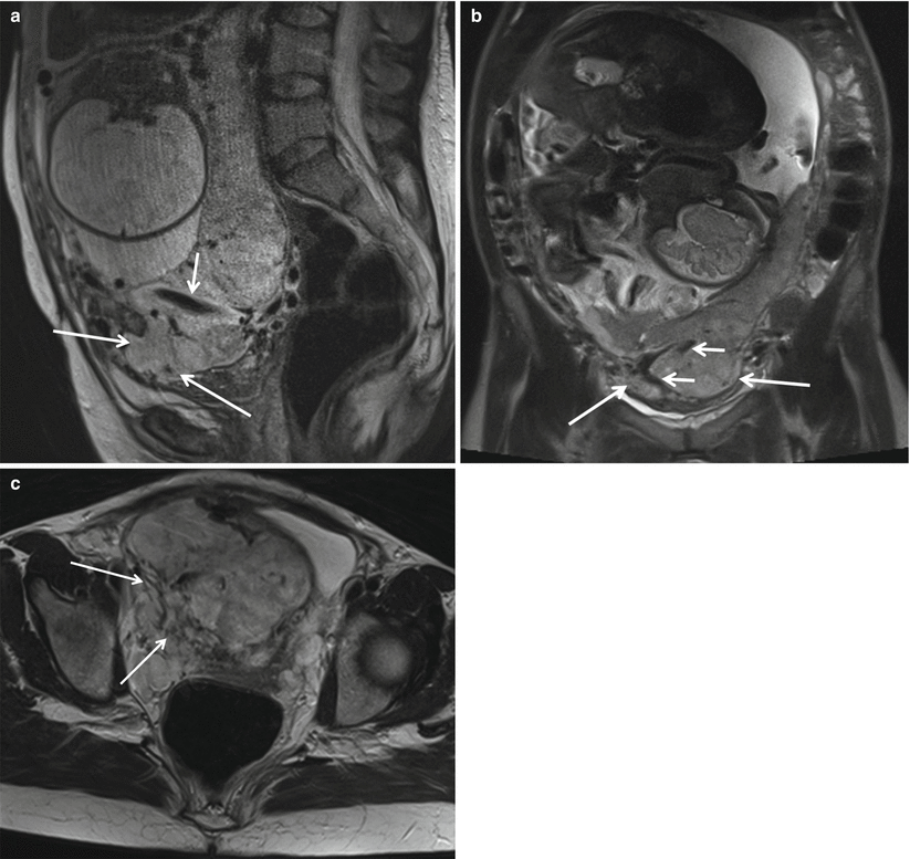
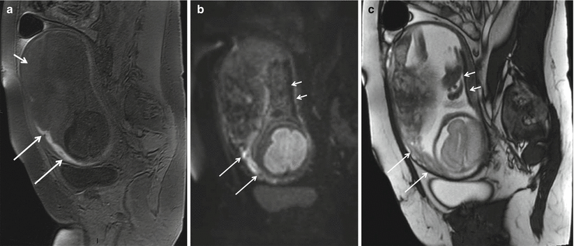
Fig. 1.1
Placenta percreta in a 28-year-old pregnant woman at 34 weeks gestation. Sagittal T2-FSE ( a ) and coronal T2-weighted HASTE images ( b ) show the bulging of the placenta ( arrows ) that invades an external layer of the bladder. Intraplacental hypointensive signal bands (newly formed vessels) are clearly seen ( short arrows ). Axial T2-weighted HASTE image ( c ) showing massive invasion of the right parametrium ( arrows ). The myometrial signal is absent in most part of the invaded area
Different studies compared the diagnostic performance of US and MRI in the diagnosis of PAD. One study retrospectively compared transvaginal US and MRI in 39 patients with abnormal placentation and showed 77 % sensitivity, 96 % specificity, 65 % PPV, and 98 % NPV for US and 88 % sensitivity, 100 % specificity, 100 % PPV, and 82 % NPV for MRI [].
Another study with 13 patients showed comparable low sensitivities of US and MRI for the diagnosis of PAD (33 % and to 38 %, respectively) [].
A recent meta-analysis reviewed 18 studies involving 1010 pregnancies at risk for invasive placentation and found no difference in the sensitivity ( P =0.24) or specificity ( P =0.91) of US and MRI for the detection of PAD; however, based on the results of a single study included in the analysis, the authors suggested that MRI should always be considered to better assess both the depth and the topography of PAD [].
In a report of 12 cases of PAD, MRI correctly gauged the depth of invasion in 3 cases underestimated by US; moreover, with reference to a standardized topographic classification that divides PADs into S1-PAD and S2-PAD (located anteriorly and posteriorly, respectively, to a plane perpendicular to the center of the so-called upper bladder axis), MRI correctly identified 3 cases of S2-PADs that were incorrectly scored as S1-PADs by US [].
1.2 Placental Abruption
Placental abruption is the leading cause of vaginal bleeding in the third trimester of pregnancy, occurring in approximately 1 % of births, and accounts for up to 25 % of perinatal deaths; it originates from a premature separation of the placenta from the uterine wall and leads to the formation of retroplacental, subchorionic, or subamniotic hematomas [].
MRI can dramatically improve the diagnosis of placental abruption. Diffusion- and T1-weighted sequences correctly identified 19 (100 %) and 18 (95 %) of 19 abruptions, with an extremely good interobserver agreement ( k =0.949) [].
The contemporary evaluation of T1-weighted, T2-weighted, and DWI images allowed the classification of hematomas as hyperacute (first few hours, intracellular oxyhemoglobin), acute (13 days, intracellular deoxyhemoglobin), early subacute (37 days, intracellular methemoglobin), and late subacute (14 days, extracellular methemoglobin) according to the paramagnetic effects of methemoglobin (Fig. ).
Fig. 1.2
Placental abruption in a 35-year-old woman at 32 weeks of gestational age presenting with history of vaginal bleeding and unremarkable US findings. Sagittal gradient echo T1-weighted image ( a ) shows a marginal anterior hematoma ( long arrows ) with blood extending into the cervix; the hematoma appears hyperintense with respect to the placenta ( short arrow ). Sagittal DWI image ( b ) shows the markedly hyperintense anterior hematoma ( long arrows ) and a hypointense hemorrhage ( short arrow ) along the posterior uterine surface. On sagittal axial true FISP ( c ), the anterior hematoma signal intensity ( long arrow ) is similar to that of the placenta, while the posterior blood collection is hypointense ( short arrow ). Therefore, the different MR sequences are complementary and are all useful for a complete characterization of existing hemorrhage
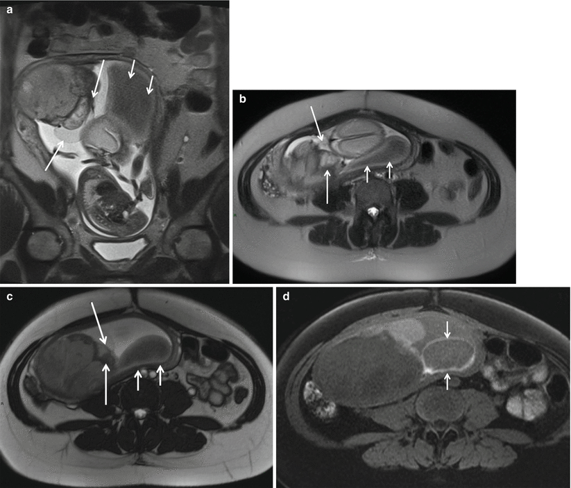
Fig. 1.3
Placental chorioangioma in patient at 24 weeks of gestational age and profuse vaginal bleeding. Coronal ( a ) and axial ( b ) T2-weighted HASTE and axial true FISP ( c ) images show an intrauterine mass peripherally located and protruding from the fetal surface of the placenta. Note the posterior hypointense subamniotic hematoma ( short arrows ). Axial T1-weighted fat-saturated ( d ) sequence shows the subamniotic hematoma ( short arrows )
1.3 MRI in the Management of Twin-To-Twin Transfusion Syndrome
Twin-to-twin-transfusion syndrome (TTTS) results from a hemodynamical imbalance of placental vascular anastomosis connecting the circulation of the two fetuses. Demise of a co-twin in a monochorionic pregnancy places the surviving twin at significant risk of death or neurologic sequelae due to severe intracranial hypoperfusion [] and is more sensitive than US in detecting ischemic injury and therefore may have a great theoretical role in the context of TTTS.
In a series of 21 cases of TTTS complicated by the demise of one twin, intracranial abnormalities including polymicrogyria, germinolytic cysts, intracranial hemorrhage, ventriculomegaly, and delayed sulcation were evidenced in seven cases by antenatal MRI; notably, in four of these seven cases, US did not score any brain abnormality, while in two of the remaining three cases, MRI showed additional findings with respect to US []. Of note, in the two abovementioned studies, MRI examinations were performed only a few days after the US examination; this limits any possible bias related to the evolutive nature of these lesions and supports the proposition that fetal brain pathology in TTTS is genuinely better imaged by MRI.