C. G. Weber M.D. - Clinical Ophthalmology - 2019
Here you can read online C. G. Weber M.D. - Clinical Ophthalmology - 2019 full text of the book (entire story) in english for free. Download pdf and epub, get meaning, cover and reviews about this ebook. year: 2014, publisher: Pacific Primary Care Software PC, genre: Romance novel. Description of the work, (preface) as well as reviews are available. Best literature library LitArk.com created for fans of good reading and offers a wide selection of genres:
Romance novel
Science fiction
Adventure
Detective
Science
History
Home and family
Prose
Art
Politics
Computer
Non-fiction
Religion
Business
Children
Humor
Choose a favorite category and find really read worthwhile books. Enjoy immersion in the world of imagination, feel the emotions of the characters or learn something new for yourself, make an fascinating discovery.

- Book:Clinical Ophthalmology - 2019
- Author:
- Publisher:Pacific Primary Care Software PC
- Genre:
- Year:2014
- Rating:4 / 5
- Favourites:Add to favourites
- Your mark:
Clinical Ophthalmology - 2019: summary, description and annotation
We offer to read an annotation, description, summary or preface (depends on what the author of the book "Clinical Ophthalmology - 2019" wrote himself). If you haven't found the necessary information about the book — write in the comments, we will try to find it.
C. G. Weber M.D.: author's other books
Who wrote Clinical Ophthalmology - 2019? Find out the surname, the name of the author of the book and a list of all author's works by series.


Clinical Ophthalmology - 2019 — read online for free the complete book (whole text) full work
Below is the text of the book, divided by pages. System saving the place of the last page read, allows you to conveniently read the book "Clinical Ophthalmology - 2019" online for free, without having to search again every time where you left off. Put a bookmark, and you can go to the page where you finished reading at any time.
Font size:
Interval:
Bookmark:
Contents:|
Tableof Contents
|
|
|
|
|
|
|
|
|
|
|
|
|
|
|
|
|
|
|
|
|
|
|
|
Primary editor Steven D. Zelko MD,Ophthalmologist.
Last updated on 28 July2018
http://clinicalmedconsult.com/
Weperform an ongoing review of most major journals, textbooks, and other online resources.Ongoing scientific information is added as the new and clinically relevantevidence is released. This is the only point of care medical reference createdfor portable electronic devices in a concise and compact format by physiciansand for physicians. The goal of this text is to bring all of the clinicallyrelevant information clinicians need during patient care directly to one'sfingertips. |
TheClinical Medicine Consult includes all 35 of our modules in the clinical medicine series in one highly integrated text . The file size of The Clinical Medicine Consult 2017v2 was 78,369kb. All of these texts are in full 64k color. Currently the totalillustrations / diagrams / color photos standsat >4,784. The iSilo version of this text comes with free quarterly updatesfor one year. Both the Kindle and iSilo versions can be read / utilized on justabout any portable device (android phone / tablet or iPhone, iPad etc), withthe appropriate reader installed (get at your app store).
Links:|
Eyeproblems constitute 2% to 3% of all primary care and emergency departmentvisits (Am Fam Physician 2016;93(12)991-998).
Despite its relatively small size, the eye isone of the most 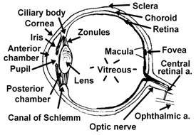 complexorgans of the human body. It is derived from neural crest cells, neuroectoderm,surface ectoderm, and mesoderm. The white tissue of the eye, the sclera, is arigid connective tissue that encloses intraocular contents except at theanterior and posterior poles, where it meets the cornea and optic nerve respectively.The conjunctiva is a diaphanous tissue that lines the inner aspect of theeyelids (tarsal conjunctiva) and reflects in the fornices to cover the sclera(bulbar conjunctiva), consists of an outer epithelial layer and underlyingloose connective tissue. As a result of its rich vascularization, theconjunctiva can demonstrate dramatic injection and edema (chemosis) in responseto irritation, allergies, or infection. Goblet cells within the conjunctivasecrete the mucous component of the tear film.
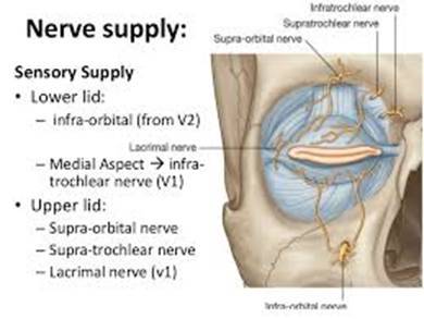
The cornea(more specifically, the air-tear film interface) is responsible for ~ twothirds of the eye's refractive power. Although like the sclera, the cornea iscomposed primarily of connective tissue (with outer epithelial and innerendothelial layers), it is transparent. This transparency is attributable tothe unique characteristic arrangement of type I collagen fibrils in a regulararray held in place by a proteoglycan ground substance. When damaged, cornealstroma undergoes further deposition of type I collagen; however, this secondaryprocess occurs without fibrillar organization, and opacification (scarring) ofcorneal stroma occurs. When the cornea is not clear, this indicates that apathologic event has occurred or is occurring.
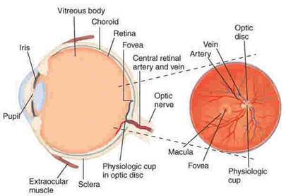 Extraocularmuscles attach to the globe underneath the conjunctiva and act as pulleys toeffect ocular rotations. Each extraocular muscle is yoked with a muscle in theother eye, causing complementary binocular function. A simplified schematicapproach to eyelid anatomy would divide the eyelid into two segments(anterior/external and posterior/internal) separated by a thin fibrousstructure, the orbital septum. The anatomic segments external to the septumconsist of skin, loose subcutaneous connective tissue in which eyelash folliclesoriginate, and orbicularis oculi muscle. Internal to the septum lie the levatormuscle, tarsus, Mller's muscle, and tarsal conjunctiva. Both segments of theeyelid contain sebaceous glands that contribute to the lipid component of thetrilayered tear film. Zeis' glands are associated with cilia (eyelashes), andmeibomian glands located within tarsus excrete secretions through orifices inthe inner eyelid margin. The nasolacrimal system consists of inflow and outflowcomponents. The lacrimal gland, a bilobed exocrine gland located under thetemporal upper eyelid, produces aqueous secretions in response to reflex andpsychogenic stimuli. Basal aqueous secretions are produced by the eccrineglands of Wolfring and Krause located adjacent to tarsus in the eyelids.Thenasolacrimal outflow tract originates at the inferior and superior puncta ofthe nasal aspect of the eyelids. They open into the superior and inferiorcanaliculi, which join to form a common canaliculus that opens into thelacrimal sac. The nasolacrimal duct exits the sac inferiorly and ends at theinferior meatus under the inferior turbinate within the nose.
Physiology of Vision:
1. Optics:Light travels in a straight line until a new medium is encountered - it maythen bend or be refracted.
a. Therefractive tissues of the eye form a convex surface.
b. Thedistance at which the bent light converges to a focal point creates threeconditions:
- Emmetropia:Focal point hits retina.
- Myopia:Focal point is in front of retina creating nearsightedness. Correctivelenses or surgery may correct refractive abnormalities.
- Hypermetropia:Focal point is behind retina, creating farsightedness.
2. Accommodation:Objects closer than six meters (20 ft.) generally have light rays that mustbe refracted greatly, thus requiring the eye to accommodate or adjust. Involvesthree major actions:
a. Lensshape: Ciliary muscle contraction regulates the shape of the lens.
Presbyopia occurswhen lens loses elasticity with age, which decreases ability to accommodate.
b. Pupilsize: Pupillary dilator muscles relax while pupillary constrictors contractto eliminate divergent light rays, making refraction easier to accomplish.
c. Eyeconvergence: Eyes turn medially to focus light on the fovea or areaof greatest visual acuity.
- Nearpoint: Minimum distance from eye that object can be focused usingaccommodation.
- Farpoint: Distance from eye that does not require accommodation; generally sixmeters (20 ft.)
20/20vision is normal focusing at 20 ft.
20/40is only focusing objects at 20 ft. that a normal eye can focus at 40 ft. 20/10 is focusing objects that a normal eye could only focus at 10 ft.
3. Photoreceptorsof the retina
a. Rods:Respond to light levels, but not color; low threshold; good in dim light(i.e., night); common in peripheral areas of retina.
b. Cones:Respond to light levels and color (red, green, blue); high threshold; goodin bright light conditions (Ex: day); common in fovea for visual acuity.
Theaverage duration of a single blink of the human eye is 0.3 seconds. The averagetime between blinks of the eye is 2.8 seconds. The average person blinks theireyes about 11,500 times per day or about 4.2 million times per year.
Theonly part of the human body that never changes size from birth to death is theeyes.
Theeye is the only part of the human body that can function at 100% ability at anymoment, day or night.
Font size:
Interval:
Bookmark:
Similar books «Clinical Ophthalmology - 2019»
Look at similar books to Clinical Ophthalmology - 2019. We have selected literature similar in name and meaning in the hope of providing readers with more options to find new, interesting, not yet read works.
Discussion, reviews of the book Clinical Ophthalmology - 2019 and just readers' own opinions. Leave your comments, write what you think about the work, its meaning or the main characters. Specify what exactly you liked and what you didn't like, and why you think so.