• Complain

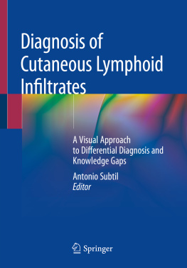
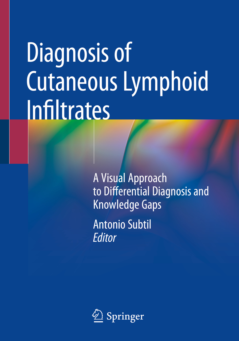
Antonio Subtil - Diagnosis of Cutaneous Lymphoid Infiltrates: A Visual Approach to Differential Diagnosis and Knowledge Gaps

Here you can read online Antonio Subtil - Diagnosis of Cutaneous Lymphoid Infiltrates: A Visual Approach to Differential Diagnosis and Knowledge Gaps full text of the book (entire story) in english for free. Download pdf and epub, get meaning, cover and reviews about this ebook. year: 2019, publisher: Springer, genre: Romance novel. Description of the work, (preface) as well as reviews are available. Best literature library LitArk.com created for fans of good reading and offers a wide selection of genres:

Romance novel Science fiction Adventure Detective Science History Home and family Prose Art Politics Computer Non-fiction Religion Business Children Humor

Choose a favorite category and find really read worthwhile books. Enjoy immersion in the world of imagination, feel the emotions of the characters or learn something new for yourself, make an fascinating discovery.

Antonio Subtil Diagnosis of Cutaneous Lymphoid Infiltrates: A Visual Approach to Differential Diagnosis and Knowledge Gaps
  • Book:
    Diagnosis of Cutaneous Lymphoid Infiltrates: A Visual Approach to Differential Diagnosis and Knowledge Gaps
  • Author:
  • Publisher:
    Springer
  • Genre:
  • Year:
    2019
  • Rating:
    3 / 5
  • Favourites:
    Add to favourites
  • Your mark:
    • 60
    • 1
    • 2
    • 3
    • 4
    • 5

Diagnosis of Cutaneous Lymphoid Infiltrates: A Visual Approach to Differential Diagnosis and Knowledge Gaps: summary, description and annotation

We offer to read an annotation, description, summary or preface (depends on what the author of the book "Diagnosis of Cutaneous Lymphoid Infiltrates: A Visual Approach to Differential Diagnosis and Knowledge Gaps" wrote himself). If you haven't found the necessary information about the book — write in the comments, we will try to find it.

This volume is the first of its kind to emphasize the visual approach in the diagnosis of cutaneous lymphoid infiltrates. Written and designed in an accessible yet highly detailed format by an expert in the field, this book bridges the knowledge gaps so often found when dealing with skin lymphomas. Complete with more than two hundred high quality images and illustrations, Diagnosis of Cutaneous Lymphoid Infiltrates offers pearls and pitfalls as well as differential diagnoses. Additionally, images are explained and decoded with the use of illustrations and analogies, proving to be an invaluable resource for pathologists, dermatologists, dermatopathologists, hematopathologists, and residents and fellows in these fields.

Antonio Subtil: author's other books


Who wrote Diagnosis of Cutaneous Lymphoid Infiltrates: A Visual Approach to Differential Diagnosis and Knowledge Gaps? Find out the surname, the name of the author of the book and a list of all author's works by series.

Diagnosis of Cutaneous Lymphoid Infiltrates: A Visual Approach to Differential Diagnosis and Knowledge Gaps — read online for free the complete book (whole text) full work

Below is the text of the book, divided by pages. System saving the place of the last page read, allows you to conveniently read the book "Diagnosis of Cutaneous Lymphoid Infiltrates: A Visual Approach to Differential Diagnosis and Knowledge Gaps" online for free, without having to search again every time where you left off. Put a bookmark, and you can go to the page where you finished reading at any time.

Light

Font size:

Reset

Interval:

Bookmark:

Make
Contents
Landmarks
Antonio Subtil Diagnosis of Cutaneous Lymphoid Infiltrates A Visual Approach - photo 1
Antonio Subtil
Diagnosis of Cutaneous Lymphoid Infiltrates A Visual Approach to Differential Diagnosis and Knowledge Gaps
Antonio Subtil Dermatology and Pathology Yale University New Haven CT USA - photo 2
Antonio Subtil
Dermatology and Pathology, Yale University, New Haven, CT, USA
ISBN 978-3-030-11652-1 e-ISBN 978-3-030-11654-5
https://doi.org/10.1007/978-3-030-11654-5
Springer Nature Switzerland AG 2019
This work is subject to copyright. All rights are reserved by the Publisher, whether the whole or part of the material is concerned, specifically the rights of translation, reprinting, reuse of illustrations, recitation, broadcasting, reproduction on microfilms or in any other physical way, and transmission or information storage and retrieval, electronic adaptation, computer software, or by similar or dissimilar methodology now known or hereafter developed.
The use of general descriptive names, registered names, trademarks, service marks, etc. in this publication does not imply, even in the absence of a specific statement, that such names are exempt from the relevant protective laws and regulations and therefore free for general use.
The publisher, the authors, and the editors are safe to assume that the advice and information in this book are believed to be true and accurate at the date of publication. Neither the publisher nor the authors or the editors give a warranty, express or implied, with respect to the material contained herein or for any errors or omissions that may have been made. The publisher remains neutral with regard to jurisdictional claims in published maps and institutional affiliations.

This Springer imprint is published by the registered company Springer Nature Switzerland AG

The registered company address is: Gewerbestrasse 11, 6330 Cham, Switzerland

Preface

The diagnosis of cutaneous lymphoid infiltrates is one of the most challenging topics of pathology, in part because this area is at the interface of several medical disciplines (dermatopathology, hematopathology, dermatology, and hematology/oncology). As a pathologist trained in both dermatopathology and hematopathology, I have noticed several knowledge gaps by dermatopathologists as well as hematopathologists. In addition, after more than a decade practicing in a referral center for skin lymphomas and participating in a busy consult service, I have witnessed recurring mistakes in the classification of cutaneous lymphoproliferative disorders and pseudolymphomas, some of which with significant clinical impact.

Both dermatology and pathology rely heavily on a visual approach to learning, developing differential diagnoses, and making final diagnoses. Pattern recognition and image decoding skills are essential elements in this process. However, most textbooks do not emphasize images nor adequately use illustrations to demystify important concepts. This disconnect is particularly critical in complicated topics, such as the correct classification of cutaneous lymphomas and pseudolymphomas. The goal of this book is to fill in important knowledge gaps utilizing a visual approach to learning. Both common and rare lymphomas are reviewed. Diagnostic pearls and pitfalls as well as differential diagnoses are covered in detail. In addition to multiple images, several tables, illustrations, and visual clues to the diagnosis are included. In order to highlight what is actually useful to make a diagnosis in real practice, genetic data that is currently only relevant at a research level is included separately in an appendix.

Beyond its usefulness to general pathologists, dermatopathologists, and hematopathologists, this book is also intended to be helpful for dermatologists, hematologists/oncologists, dermatopathology fellows, hematopathology fellows, pathology residents, and dermatology residents.

I would like to acknowledge and thank my mother, Cezina; my late father, Antonio; and my sisters, Adriane and Cristiane, for their lifelong support. Thank you to Larry Gibson, Ifty Ahmed, Jeannine Holden, Peter Heald, Jennifer McNiff, Rick Edelson, Mark Pittelkow, Jean Bolognia, Earl Glusac, Christine Ko, Anjela Galan, Francine Foss, Mike Girardi, Peggy Myung, Shawn Cowper, Irwin Braverman, Kal Watsky, Alistair Robson, Joan Guitart, Christiane Querfeld, Jaqueline Junkins-Hopkins, Catherine Stefanato, Russell Fiorella, Kamani Lankachandra, Edward Gutmann, Carole McArthur, Roberto Miranda, Jamie Skrade, Peter Kragel, Karen Mann, David Jaye, Shiyong Li, Rich Antaya, Jae Choi, Jennifer Choi, Bob Tigelaar, Mary Tomayko, Kacie Carlson, Ann Putio, Jean Saley, Sasha Finn, Carey Storan, Sikina Rossi, Jinah Kim, Uma Sundram, Youn Kim, Eleanor Knopp, Phil Shapiro, Rossitza Lazova, Steven Billings, Alina Bridges, Margot Peters, Diane Pierson, Adriane Brito, Lola Pettinicchio, Ricardo Macarenco, Jane Grant-Kels, Sharon Weiss, Tony Neto, Stephen Joyner, John Napoli, Gauri Panse, Brian Poligone, Phil LeBoit, Marcus Bosenberg, MaryAnn Ackerman, Debbie Williams, Terri Borrowman, Andrea Harris, Cristina Ishihama, Anini Group (Kelia, Adriane, Thays, Ricardo, Claudiney, Lusmaia, Greice, Maria Cristina, Patrycia, Claudio, Renato), Thanila Macedo, Marilia Ribeiro, Lara Quirino, Lucelia Badan, Celia Revilandia, Scott Freese, Jon Talbert, Marie Valdez, Nancy Talbert, Jack Gryder, Alice Gryder, Leigh Ann Homb, and Norman Homb. Thank you also to Lorraine Coffey, Rebekah Amos, and Samantha Lonuzzi at Springer for their support and outstanding skills. Lastly, thank you to my husband, Nathan, for turning the television volume down so I could write.

Antonio Subtil
New Haven, CT, USA
Contents
Part IThe Basics
Part IIHistomorphologic Differential Diagnoses (DDx)
Part IIISpecial Techniques
Part IVDiagnostic Entities
Part I The Basics
Springer Nature Switzerland AG 2019
Antonio Subtil Diagnosis of Cutaneous Lymphoid Infiltrates https://doi.org/10.1007/978-3-030-11654-5_1
1. Clinical Pathological Correlation
Antonio Subtil
(1)
Dermatology and Pathology, Yale University, New Haven, CT, USA
Antonio Subtil
Keywords
Skin Lymphoma Clinical Histopathology Correlation

Careful correlation of clinical and histopathologic findings is essential to correctly classify cutaneous lymphoid infiltrates. Skin lymphomas would be more appropriately viewed as syndromes, in which a constellation of clinical as well as pathologic changes is needed for each diagnosis. Any given finding cannot be used in a vacuum to make a diagnosis. Ignoring either side of this clinicopathologic equation will often result in misclassification.

A similar histomorphologic pattern is often seen in different cutaneous lymphomas, often with markedly different clinical behaviors. Therefore, depending on the clinical presentation, different diagnoses will be made for similar histopathologic findings (Fig. ). However, MF would present with large, chronic patches and/or plaques on sun-protected skin, while the clinical picture of LyP is of small, self-regressing papules. Epidermotropism may also be seen in aggressive cytotoxic lymphomas, such as primary cutaneous CD8+ aggressive epidermotropic cytotoxic T-cell lymphoma and cutaneous gamma-delta T-cell lymphoma .
Next page
Light

Font size:

Reset

Interval:

Bookmark:

Make

Similar books «Diagnosis of Cutaneous Lymphoid Infiltrates: A Visual Approach to Differential Diagnosis and Knowledge Gaps»

Look at similar books to Diagnosis of Cutaneous Lymphoid Infiltrates: A Visual Approach to Differential Diagnosis and Knowledge Gaps. We have selected literature similar in name and meaning in the hope of providing readers with more options to find new, interesting, not yet read works.


Reviews about «Diagnosis of Cutaneous Lymphoid Infiltrates: A Visual Approach to Differential Diagnosis and Knowledge Gaps»

Discussion, reviews of the book Diagnosis of Cutaneous Lymphoid Infiltrates: A Visual Approach to Differential Diagnosis and Knowledge Gaps and just readers' own opinions. Leave your comments, write what you think about the work, its meaning or the main characters. Specify what exactly you liked and what you didn't like, and why you think so.