Ernesto Bonifazi - Differential diagnosis in pediatric dermatology
Here you can read online Ernesto Bonifazi - Differential diagnosis in pediatric dermatology full text of the book (entire story) in english for free. Download pdf and epub, get meaning, cover and reviews about this ebook. year: 2013, publisher: Springer Verlag, genre: Detective and thriller. Description of the work, (preface) as well as reviews are available. Best literature library LitArk.com created for fans of good reading and offers a wide selection of genres:
Romance novel
Science fiction
Adventure
Detective
Science
History
Home and family
Prose
Art
Politics
Computer
Non-fiction
Religion
Business
Children
Humor
Choose a favorite category and find really read worthwhile books. Enjoy immersion in the world of imagination, feel the emotions of the characters or learn something new for yourself, make an fascinating discovery.

- Book:Differential diagnosis in pediatric dermatology
- Author:
- Publisher:Springer Verlag
- Genre:
- Year:2013
- Rating:5 / 5
- Favourites:Add to favourites
- Your mark:
Differential diagnosis in pediatric dermatology: summary, description and annotation
We offer to read an annotation, description, summary or preface (depends on what the author of the book "Differential diagnosis in pediatric dermatology" wrote himself). If you haven't found the necessary information about the book — write in the comments, we will try to find it.
Ernesto Bonifazi: author's other books
Who wrote Differential diagnosis in pediatric dermatology? Find out the surname, the name of the author of the book and a list of all author's works by series.


Differential diagnosis in pediatric dermatology — read online for free the complete book (whole text) full work
Below is the text of the book, divided by pages. System saving the place of the last page read, allows you to conveniently read the book "Differential diagnosis in pediatric dermatology" online for free, without having to search again every time where you left off. Put a bookmark, and you can go to the page where you finished reading at any time.
Font size:
Interval:
Bookmark:
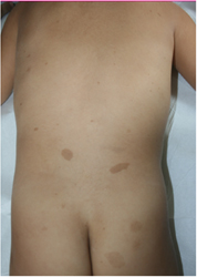
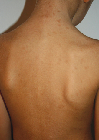
1. Neurofibromatosi | 2. Macular Mastocytosis | |
|---|---|---|
Definition | Autosomal dominant inherited disease, initially characterized by caf au lait spots. | Benign proliferation of dermal mastocytes clinically characterized by brownish maculae. |
Family history | Present in 30% of cases []. | Present in 3% of cases []. |
Time of onset | Often present at birth or in the first months. | Mastocytosis is sometimes present at birth. It becomes evident in the first months of life. |
Sites involved | Anywhere on the skin. | Mainly on the chest. |
Darier sign | Absent. | Present, although barely evident due to the paucity of mastocytes. |
Involvement of the groin region and axillary folds | Frequent. | Rare. |
Lesion size | Highly variable, even within the same subject; larger lesions can exceed 5 cm; the smallest may measure less than 5 mm. | Rather uniform, usually 12 cm in diameter. |
Lesion outline | Clearly defined. | Blurred. |
Development | New lesions, usually smaller, appear with age. Pre-existing lesions usually persist unchanged. | No new lesions appear after the first year. Pre-existing lesions disappear in several years. |
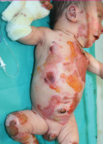
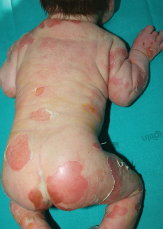
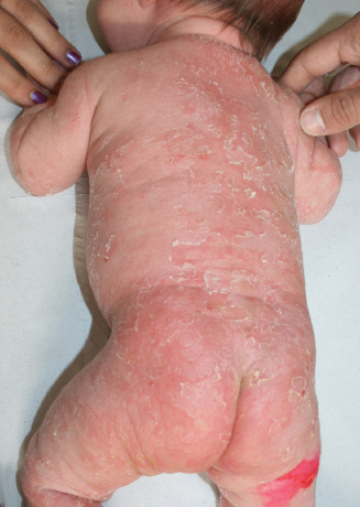
1. Recessive Dystrophic Epidermolysis Bullosa | 2. Bullous Congenital Ichthyosiform Erythroderma | |
|---|---|---|
Definition | Inherited disease due to a collagen defect, with consequent post-traumatic blisters and dystrophic scars. | Inherited disease due to a keratin defect, with consequent blisters and scaling erythroderma. |
Heredity | Autosomal recessive. | Autosomal dominant. |
Blood blisters | Often present. | Lacking. |
Blister distribution | Hands, feet, elbows, knees. | Trunk, limbs. |
Mouth blisters | Often present. | Lacking. |
Generalized erythema | Lacking. | Present. |
Healing of blisters | Slow, with milia and scarring. | Rapid, without scarring. |
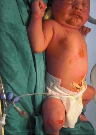
1. Aplasia Cutis Congenita | 2. Sebaceous Nevus in the Newborn | |
|---|---|---|
Definition | Congenital, localized absence of epidermis, dermis and sometimes subcutaneous tissue, which heals with scarring. | Nevus with prominent involvement of the sebaceous glands. |
Number of lesions | Usually single, sometimes multiple. | Usually only one. |
Lesion morphology | Eroded or already scarred at birth. In the latter, the surface is smooth. | Granular. |
Clinical course |
Font size:
Interval:
Bookmark:
Similar books «Differential diagnosis in pediatric dermatology»
Look at similar books to Differential diagnosis in pediatric dermatology. We have selected literature similar in name and meaning in the hope of providing readers with more options to find new, interesting, not yet read works.
Discussion, reviews of the book Differential diagnosis in pediatric dermatology and just readers' own opinions. Leave your comments, write what you think about the work, its meaning or the main characters. Specify what exactly you liked and what you didn't like, and why you think so.