CHAPTER 4
MCAT EXAM PRACTICE Test 2
CHAPTER SUMMARY
This is the second of two practice MCAT exams.
T he exam is organized in four sections:
Biological and Biochemical Foundations of Living Systems
Chemical and Physical Foundations of Biological Systems
Psychological, Social, and Biological Foundations of Behavior
Critical Analysis and Reasoning Skills
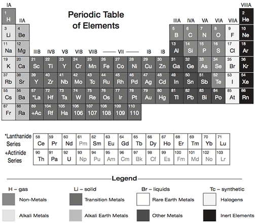
(Note: you will have access to the Periodic Table of Elements during the exam.)
This practice test is modeled on the content, format, and length of the official MCAT exam. At the beginning of each section youll find the number of questions and the time allotted. We recommend that you take the test under timed conditions so you can get an accurate assessment of how you might do on test day and determine the pace youll need to work at.
Use your results on each section of the exam to determine your strengths and weaknesses and guide your study. Good luck!

Part 1: Biological and Biochemical Foundations of Living Systems
95 Minutes
59 Questions
Use the following passage to answer questions 1 through 4.
Very few proteins carry out their functions in the cell as unmodified folded polypeptides. Nearly all proteins undergo post-translational modifications (PTMs) in various cellular compartments, including the cytosol, Golgi apparatus, and endoplasmic reticulum. One common modification type is proteolytic cleavage, which involves irreversible processing of a polypeptide by proteases. Another major category of PTMs is covalent modification of specific amino acid residues. Among the many examples of these modifications are phosphorylation, acetylation, methylation, and glycosylation. There is evidence that every amino acid residue can be decorated with one or more of these modification groups. The specificity of this type of PTM is partially determined by the pattern of amino acids surrounding the target residue. Because PTMs affect myriad properties of proteins, including stability, localization, and interaction with other proteins, there has been intense effort in biological research to characterize the PTMs of individual proteins and protein populations in the cell. The influence of PTMs on protein function is illustrated by the example of p53, a canonical tumor suppressor protein. The levels of p53 in mammalian cells are tightly regulated by a modification called ubiquitination. Under normal conditions, the addition of multiple ubiquitin groups to p53 results in the sequestration of the protein into the 26S proteasome complex and its consequent clearance from the cell. However, when the cell encounters stress, such as DNA damage or the activation of an oncogene, another pattern of PTM confers the stabilization of p53. Under these conditions, ubiquitination is suppressed, and p53 is instead acetylated and phosphorylated; both of these modifications stabilize the protein. P53 primarily functions as a transcriptional activator or repressor, and its target genes are involved in cell cycle arrest, apoptosis, and DNA repair. It accomplishes these functions in homotetrameric complexes, and phosphorylation of p53 has additionally been demonstrated to increase the complexs sequence-specific binding to DNA. PTMs are dynamic; phosphorylation, which often occurs in the cytosol, is controlled by kinase enzymes, whereas removal of phosphate groups is carried out by another set of enzymes called phosphatases. Similarly, while histone acetyltransferases add acetyl groups to p53, histone deacetylases remove them. P53 has also been shown to undergo additional PTMs: neddylation, which could repress its transactivator activity; sumoylation, which could act like ubiquitination; glycosylation; and ribosylation. The importance of p53 in cancer formation is illustrated by the fact that more than 20,000 mutations in the protein have been found in various cancer types. Many mutations lie in the p53 DNA binding domain and give rise to oncogenic properties. Additionally, mutations often result in increased phosphorylation and acetylation and, thus, stabilization of these oncogenic forms of p53.
Which of the following post-translational modifications is most likely to mark a protein for degradation?
a. Monoubiquitination
b. Polyubiquitination
c. Acetylation
d. Neddylation
Which of the following is a property of the cellular compartment where protein phosphorylation often occurs?
a. It is aqueous.
b. It is usually only found in eukaryotes.
c. It has a pH of 6.5.
d. It precipitates away from the other cellular compartments by high-speed centrifugation.
Which of the following is NOT a true similarity between covalent modifications and proteolytic cleavage?
a. Both processes affect the cellular function of proteins.
b. Both processes are controlled by enzymes.
c. Both processes are reversible.
d. Both processes affect protein localization.
Which method would likely NOT allow for the detection of different modification states of a protein?
a. Deletion of an amino acid that bears the modification
b. Mass spectrometry
c. 2D gel electrophoresis
d. Restriction enzyme digest
Which of the following amino acids is typically modified with a phosphate group?
a. Proline
b. Serine
c. Tryptophan
d. Valine
Use the following passage to answer questions 6 through 10.
Glucokinase possesses several properties that make it an attractive target for type 2 diabetes therapy, and several activators of the enzyme are currently being developed as potential drug candidates. Although there are numerous treatment options for type 2 diabetes, none of them allow for long-lasting control of blood glucose in most patients. Glucokinase, also known as hexokinase IV, is expressed in the liver and mediates the clearance of glucose from the blood through at least two different mechanisms. First, it catalyzes the phosphorylation of glucose to glucose 6-phosphate. Other forms of hexokinase (hexokinase I, II, III) catalyze the same reaction in other cells of the body, such as muscle cells. Second, glucokinase controls the levels of glucose by regulating the conversion of glucose to the storage form of glycogen. There is also evidence that glucokinase acts as a glucose sensor in pancreatic cells, where it is differentially expressed in response to glucose. These research observations led to speculation that glucokinase might be an important determinant of the diabetes phenotype. Indeed, family studies in the 1990s revealed that mutations associated with reduced glucokinase activity were associated with a form of diabetes known as maturity onset diabetes of the young (MODY). Furthermore, there is evidence that inactivating mutations act in a dose-dependent fashion: mutations in a single allele are linked to mild hyperglycemia, whereas homozygous mutations are linked to severe permanent diabetes in the neonatal stages. On the other hand, activating mutations have been found to cause hypoglycemia. The discovery of an allosteric site in glucokinase outside of its active site paved the way for the development of enzymatic activators starting in the early 2000s. In a recent study of patients with type 2 diabetes, one such activator was found to lower blood glucose. To date, numerous glucokinase activators have been developed, and their amino acid sequence may help lead to the identification of endogenous activators of the enzyme. In contrast, there is a physiologic negative allosteric effector of glucokinase, called fructose 6-phosphate, which is a product of the glycolytic pathway downstream of glucose 6-phosphate. Fructose 6-phosphate causes glucokinase to bind more tightly to a regulatory protein that sequesters the enzyme in the nucleus when blood glucose is low. However, when blood glucose is in the normal range, glucokinase dissociates from this regulatory protein, helping to keep the levels of glucose in the appropriate physiological range.
Next page


