Robbins and Cotran Pathology Flash Cards
SECOND EDITION
Edward C. Klatt MD
Professor of Pathology, Department of Biomedical Sciences, Mercer University School of Medicine, Savannah, Georgia
Director, Biomedical Education Program, Mercer University School of Medicine, Savannah, Georgia
Richard N. Mitchell MD, PhD
Professor of Pathology, Harvard Medical School and Health Sciences and Technology, Brigham and Women's Hospital, Boston, Massachusetts
Director, Human Pathology, HarvardMIT Division of Health Sciences and Technology, Boston, Massachusetts
Staff Pathologist, Brigham and Women's Hospital, Boston, Massachusetts

Table of Contents
Copyright

1600 John F. Kennedy Blvd.
Ste. 1800
Philadelphia, PA 19103-2899
ROBBINS AND COTRAN PATHOLOGY FLASH CARDS
SECOND EDITION ISBN: 978-0-323-35222-2
Copyright 2016, 2010 by Saunders, an imprint of Elsevier Inc.
All rights reserved. No part of this publication may be reproduced or transmitted in any form or by any means, electronic or mechanical, including photocopying, recording, or any information storage and retrieval system, without permission in writing from the publisher.
Permissions may be sought directly from Elsevier's Rights Department: phone: (+1) 215 239 3804 (US) or (+44) 1865 843830 (UK); fax: (+44) 1865 853333; e-mail: .
NoticeNeither the publisher nor the authors assume any responsibility for any loss or injury and/or damage to persons or property arising out of or related to any use of the material contained in this book. It is the responsibility of the treating practitioner, relying on independent expertise and knowledge of the patient, to determine the best treatment and method of application for the patient.The Publisher
Executive Content Strategist: William Schmitt
Content Development Specialist: Amy Meros
Publishing Services Manager: Anne Altepeter
Project Manager: Louise King
Design Manager: Xiaopei Chen
Printed in China
Last digit is the print number: 9 8 7 6 5 4 3 2 1

Acknowledgments
The Flash Cards represent the work of many people. Here are two of them.
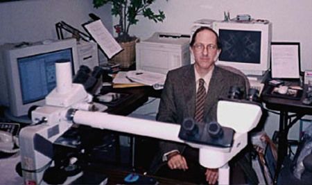

1. Who are these guys?
2. How will I use these cards?
3. Can I learn everything I need to know from these cards?
4. Who should I thank for putting all this together (besides the authors)?
5. What is the ultimate answer? Name the source.
Answers
1. At left is Edward C. Klatt, MD, professor of Pathology, Department of Biomedical Sciences, Mercer University School of Medicine; the other guy is Rick Mitchell, MD, PhD, professor of Pathology and Health Sciences and Technology, Harvard Medical School.
2. Each card has two sides. Each side includes a vignette with text, image, and questions. Flip the card for the answers to questions on the opposite side. Both card sides illustrate the same or related disease process. This style effectively doubles your learning opportunities.
3. No, of course not. By design these flash cards only hit the highlights and scratch the surface. Use them to refresh concepts, but be curious and not content to let your knowledge end there. There's so much out there to learn. Repeat after us: I will enthusiastically seek knowledge in dedicating myself to the care of others at the highest level.
4. Thank the authors of Robbins and Cotran Pathologic Basis of Disease and Basic Pathology, the texts that serve as the primary source authority of information for the flash cards. Also, thank the people at Elsevier, Inc., including William Schmitt, executive content strategist, who had the inspiration for this project and guided it to completion. Finally, thank Amy Meros, content development specialist, who provided invaluable service (and infinite patience) to move the project from concept to reality. In addition, we are indebted to our designer, XiaoPei Chen, and the project manager, Louise King, for their assistance.
5. Love is the healer for all our ills (Rumi of Balkh, 12071273)from his many sources of inspiration and enlightenmentnot on your exams, but essential to good medicine.
Preface
The second edition of Robbins and Cotran Pathology Flash Cards is designed to reinforce the learning that begins with the study of other works in the Robbins family of textbooks, as well as in other clinical and basic science disciplines. The learning styles of our user base of students are continually evolving, and we now incorporate a variety of resources to support them. The Robbins list of titles continues to expand to support such a diversity of learning modalities. These flash cards are designed to stimulate recall and reinforce concepts in pathophysiology. Use the cards to consolidate your learning and inspire further study.
UNIT I
General Pathology
OUTLINE
Cellular Responses to Stress and Toxic Insult
Adaptation, Injury, and Death
1.1 Side A
(PBD9: 36; BP9: 4)
Questions
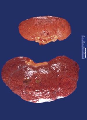
A 70-year-old woman has had a blood pressure of 160/105 mm Hg for many years. Abdominal ultrasound shows the decreased size of one kidney.
1. What gross morphologic description applies to the abnormal kidney?
2. What is the likely cause of this finding?
3. How could this affect the patient's renal function?
4. What cellular organelle plays a major role in this process?
5. What cellular protein processing pathway is involved?
Answers: Side B
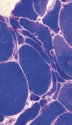
1. Myocytecellular atrophyis present as a result oflysosomal autophagyand increasedproteasomal degradation.
2. Small angulated fibers with occasional central nuclei are grouped together.
3. Skeletal muscle fibers in a motor unit are randomly enervated; nerve injury initially leads to scattered myocyte atrophy within any given motor unit. After one nerve is injured, however, an adjacent neuron can branch and reinnervate denervated myocytes. If that neuron is now injured, the result isgroup atrophyof myocytes.
1.1 Side B
(PBD9: 36; BP9: 4)
Questions
A 55-year-old man has repeated trauma to his upper arms from operating a jackhammer. He now has hand and forearm weakness. A skeletal muscle biopsy specimen reveals the pattern shown at the right.
1. What is the microscopic description of these myocytes?
2. What features support the diagnosis?
3. Why are smaller angulated fibers grouped together?
