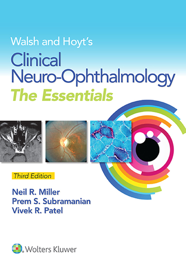
Walsh and Hoyts
Clinical Neuro-Ophthalmology
The Essentials
Third Edition
Neil R. Miller, MD
Professor of Ophthalmology, Neurology, and Neurosurgery
Frank B. Walsh Professor of Neuro-Ophthalmology
Director, Neuro-Ophthalmology and Orbit Units
Wilmer Eye Institute
Johns Hopkins Hospital
Baltimore, Maryland
Prem S. Subramanian, MD
Associate Professor of Neuro-Ophthalmology
Wilmer Eye Institute
Johns Hopkins Hospital
Baltimore, Maryland
Vivek R. Patel, MD
Wilmer Eye Institute
Johns Hopkins Hospital
Baltimore, Maryland

Acquisitions Editor: Kel MacGowan
Senior Product Development Editor: Emilie Moyer
Editorial Assistant: Diane Solomon
Marketing Manager: Stephanie Kindlick
Production Project Manager: David Orzechowski
Design Coordinator: Holly McLaughlin
Art Director/Media: Freddie Patane
Manufacturing Coordinator: Beth Welsh
Prepress Vendor: Aptara, Inc.
3rd edition
Copyright 2016 Wolters Kluwer
2nd edition Copyright 2008 Wolters Kluwer Health/Lippincott Williams & Wilkins. 1st edition Copyright 1999 Williams & Wilkins. All rights reserved. This book is protected by copyright. No part of this book may be reproduced or transmitted in any form or by any means, including as photocopies or scanned-in or other electronic copies, or utilized by any information storage and retrieval system without written permission from the copyright owner, except for brief quotations embodied in critical articles and reviews. Materials appearing in this book prepared by individuals as part of their official duties as U.S. government employees are not covered by the above-mentioned copyright. To request permission, please contact Wolters Kluwer at Two Commerce Square, 2001 Market Street, Philadelphia, PA 19103, via email at (products and services).
9 8 7 6 5 4 3 2 1
Printed in China
978-1-4511-9447-0
1-45119-447-1
Library of Congress Cataloging-in-Publication Data
available upon request
This work is provided as is, and the publisher disclaims any and all warranties, express or implied, including any warranties as to accuracy, comprehensiveness, or currency of the content of this work.
This work is no substitute for individual patient assessment based upon healthcare professionals examination of each patient and consideration of, among other things, age, weight, gender, current or prior medical conditions, medication history, laboratory data and other factors unique to the patient. The publisher does not provide medical advice or guidance and this work is merely a reference tool. Healthcare professionals, and not the publisher, are solely responsible for the use of this work including all medical judgments and for any resulting diagnosis and treatments.
Given continuous, rapid advances in medical science and health information, independent professional verification of medical diagnoses, indications, appropriate pharmaceutical selections and dosages, and treatment options should be made and healthcare professionals should consult a variety of sources. When prescribing medication, healthcare professionals are advised to consult the product information sheet (the manufacturers package insert) accompanying each drug to verify, among other things, conditions of use, warnings and side effects and identify any changes in dosage schedule or contraindications, particularly if the medication to be administered is new, infrequently used or has a narrow therapeutic range. To the maximum extent permitted under applicable law, no responsibility is assumed by the publisher for any injury and/or damage to persons or property, as a matter of products liability, negligence law or otherwise, or from any reference to or use by any person of this work.
LWW.com
To Our Patients: Past, Present, and Future
PREFACE
This third edition of Walsh and Hoyts Clinical Neuro- Ophthalmology: The Essentials, is borne from the same need that motivated the development of its 1st edition 15 years agoto provide a thorough, yet much more concise complement to the 3-volume sixth edition of the comprehensive parent text, Walsh and Hoyts Clinical Neuro-Ophthalmology. We are encouraged by the extent to which our readers have embraced the first two editions; however, we felt that given the tremendous advances that have occurred over the past decade, it was time for an updated, refined version while preserving its brevity and readability. In particular, we have emphasized the role of modern imaging techniques such as OCT, high resolution CT/MRI, and multifocal ERG in patients with known and presumed neuro-ophthalmologic disorders. Diagrams and tables have been enhanced wherever possible, and the vast majority of photos are now in color. Coverage of complex eye movement disorders has been greatly enriched by the inclusion of videos available online.
In this new edition, we have incorporated new information, both text and illustrations, within the pre-existing organization and content of its predecessors. Five sectionsthe afferent and efferent visual systems, disorders of the pupil, disorders of the eyelids, and non-organic disordersprovide the framework for 23 chapters. As we recognize that the way in which we acquire information is constantly evolving and that the speed with which information changes also is increasing, we have not included any references but trust that the reader interested in more detailed information will find it online. Nevertheless, we believe that there continues to be a need for knowledge of the fundamentals of neuro-ophthalmology, allowing the reader to integrate more effectively this knowledge with information acquired from the multitude of sources available.
We sincerely hope we have accomplished our goal of enhancing our readers understanding and appreciation of the fascinating world of neuro-ophthalmology.
CONTENTS
SECTION I
THE AFFERENT VISUAL SYSTEM
1 Examination of the Visual Sensory System
Despite continuous advances in neuroimaging and other new techniques, the examination of the afferent visual sensory system is still the core of the neuro-ophthalmologic examination. This chapter describes the most common subjective and objective testing parameters used in the afferent visual system examination.
Evaluation of the afferent system begins with a thorough medical history, followed by an ophthalmologic examination, including assessment of best-corrected or at least pinhole visual acuity at distance and near, color vision, visual fields, anterior segments (including the media), vitreous, intraocular pressure (IOP), and appearance of the optic discs, retinas (especially the macula), and retinal vessels. At the completion of the examination, one should have an idea as to the structure involved in the patients visual sensory difficulties or at least have a differential diagnosis. If the diagnosis remains unclear, a number of ancillary tests are available, including ocular imaging and electrophysiologic procedures, that should lead to the correct diagnosis.


