Table of Contents
List of Tables
- Chapter 1
List of Illustrations
- Chapter 1
- Chapter 2
- Chapter 3
- Chapter 4
- Chapter 5
- Chapter 6
- Chapter 7
- Chapter 8
- Chapter 9
- Chapter 10
- Chapter 11
Guide
Pages
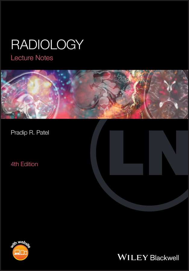
Radiology
Lecture Notes
Pradip R. Patel
Consultant Radiologist
Kingston Hospital
London, UK
Fourth Edition
First edition awarded BMA Book Award

This edition first published 2020 2020 by John Wiley & Sons Ltd
Edition History [3e, 2011]
All rights reserved. No part of this publication may be reproduced, stored in a retrieval system, or transmitted, in any form or by any means, electronic, mechanical, photocopying, recording or otherwise, except as permitted by law. Advice on how to obtain permission to reuse material from this title is available at http://www.wiley.com/go/permissions.
The right of Pradip R. Patel to be identified as the author of editorial work has been asserted in accordance with law.
Registered Office(s)
John Wiley & Sons, Inc., 111 River Street, Hoboken, NJ 07030, USA
John Wiley & Sons Ltd, The Atrium, Southern Gate, Chichester, West Sussex, PO19 8SQ, UK
Editorial Office
9600 Garsington Road, Oxford, OX4 2DQ, UK
For details of our global editorial offices, customer services, and more information about Wiley products visit us at www.wiley.com.
Wiley also publishes its books in a variety of electronic formats and by printondemand. Some content that appears in standard print versions of this book may not be available in other formats.
Limit of Liability/Disclaimer of Warranty
The contents of this work are intended to further general scientific research, understanding, and discussion only and are not intended and should not be relied upon as recommending or promoting scientific method, diagnosis, or treatment by physicians for any particular patient. In view of ongoing research, equipment modifications, changes in governmental regulations, and the constant flow of information relating to the use of medicines, equipment, and devices, the reader is urged to review and evaluate the information provided in the package insert or instructions for each medicine, equipment, or device for, among other things, any changes in the instructions or indication of usage and for added warnings and precautions. While the publisher and authors have used their best efforts in preparing this work, they make no representations or warranties with respect to the accuracy or completeness of the contents of this work and specifically disclaim all warranties, including without limitation any implied warranties of merchantability or fitness for a particular purpose. No warranty may be created or extended by sales representatives, written sales materials or promotional statements for this work. The fact that an organization, website, or product is referred to in this work as a citation and/or potential source of further information does not mean that the publisher and authors endorse the information or services the organization, website, or product may provide or recommendations it may make. This work is sold with the understanding that the publisher is not engaged in rendering professional services. The advice and strategies contained herein may not be suitable for your situation. You should consult with a specialist where appropriate. Further, readers should be aware that websites listed in this work may have changed or disappeared between when this work was written and when it is read. Neither the publisher nor authors shall be liable for any loss of profit or any other commercial damages, including but not limited to special, incidental, consequential, or other damages.
Library of Congress CataloginginPublication Data
Names: Patel, Pradip, author.
Title: Lecture notes. Radiology / Pradip Patel, Kingston Hospital, Woodlands, Kingston, UK.
Description: Fourth edition. | Hoboken : Wiley, 2020. | Series: Lecture notes | Includes bibliographical references and index.
Identifiers: LCCN 2020001869 (print) | LCCN 2020001870 (ebook) | ISBN 9781119550341 (paperback) | ISBN 9781119550334 (adobe pdf) | ISBN 9781119550372 (epub)
Subjects: LCSH: Radiography, MedicalOutlines, syllabi, etc.
Classification: LCC RC78.17 .P385 2010 (print) | LCC RC78.17 (ebook) | DDC 616.07/572dc23
LC record available at https://lccn.loc.gov/2020001869
LC ebook record available at https://lccn.loc.gov/2020001870
Cover Design: Wiley
Cover Image: Courtesy of Pradip R. Patel; piranka/Getty Images
List of contributors
Dr Patricia Marin Crespo, Consultant Radiologist, Kingston Hospital, UK
Dr Aghi Srikanthan, Consultant Radiologist, Kingston Hospital, UK
Dr Vin Majuran, Consultant Radiologist, Kingston Hospital, UK
Dr Sarah Evans, Consultant Radiologist, Kingston Hospital, UK
Dr Ramesh Yella, Consultant Radiologist, Kingston Hospital, UK
Dr Nevin Wijisekera, Consultant Radiologist, Kingston Hospital, UK
Dr Nim Mangat, Consultant Radiologist, Kingston Hospital, UK
Dr Pradip R. Patel, Consultant Radiologist, Kingston Hospital, UK
Dr Bijan Heydati, Consultant Radiologist, Kingston Hospital, UK
Dr Charlotte Whittaker, Consultant Radiologist, Kingston Hospital, UK
Dr Derek SvastiSalee, Consultant Radiologist, Kingston Hospital, UK
Dr Pradip R. Patel, Consultant Radiologist, Kingston Hospital, UK
Dr Pradip R. Patel, Consultant Radiologist, Kingston Hospital, UK
About the companion website
This book is accompanied by a companion website:
www.wiley.com/go/patel/radiology
The website includes:
- Selfassessment exercises to accompany each book chapter
- Chapter based exercises with further examples and images
Note: The website should be live by December 2020.
Introduction
Recent technological advances have produced a bewildering array of complex imaging techniques and procedures. The basic principle of imaging, however, remains the anatomical demonstration of a particular region and related abnormalities, the principal imaging modalities being:
- plain Xrays: utilizes a collimated Xray beam to image the chest, abdomen, skeletal structures, etc.;
- fluoroscopy: a continuous Xray beam produces a moving image to monitor examinations such as barium meals, barium enemas, etc.;
- ultrasound (US): employs highfrequency sound waves to visualize structures in the abdomen, pelvis, neck and peripheral soft tissues;
- computed tomography (CT): obtains crosssectional computerized densities and images from an Xray beam/detector system;
- magnetic resonance imaging (MRI): exploits the magnetic properties of hydrogen atoms in the body to produce images;
- nuclear medicine (NM): acquires functional as well as anatomical details by gamma radiation detection from injected radioisotopes.
Contrast media
Contrast agents are substances that assist visualization of some structures during the above techniques, working on the basic principle of Xray absorption, thereby preventing their transmission through the patient. The most commonly used are barium sulphate to outline the gastrointestinal tract, and organic iodine preparations; the latter widely used intravenously in CT for vascular and organ enhancement. Contrast agents can also be introduced into specific sites, for example:
Next page

