Copyright 2003, 2008 by Chris Jarmey. All rights reserved. No portion of this book, except for brief review, may be reproduced, stored in a retrieval system, or transmitted in any form or by any meanselectronic, mechanical, photocopying, recording, or otherwisewithout the written permission of the publisher. For information, contact Lotus Publishing or North Atlantic Books. First published in 2003.
British Library Cataloguing in Publication Data A CIP record for this book is available from the British Library
eBook ISBN: 978-1-58394-731-9
ISBN 978 1 905367 11 5 (Lotus Publishing)
Trade Paperback ISBN 978 1 55643 719 9 (North Atlantic Books)
The Library of Congress has cataloged the first edition as follows: Jarmey, Chris.
The concise book of muscles / Chris Jarmey.
p.; cm. ill.
Includes bibliographical references and index.
1. ill.
Includes bibliographical references and index.
1.
MusclesHandbooks, manuals, etc.
[DNLM: 1. MusclesinnervationAtlases. 2.
MusclesinnervationHandbooks. 3. Anatomy, RegionalAtlases. 4.
Anatomy, RegionalHandbooks. 5. Musclesanatomy & histologyAtlases.
6. Musclesanatomy & histologyHandbooks. WE 17 J37c 2003] I. Title.
QP321.J43 2003
612.74dc21
2002155580 v3.1
Contents
About this Book
This book is designed in quick reference format to offer useful information about the main skeletal muscles that are central to sport, dance and exercise.
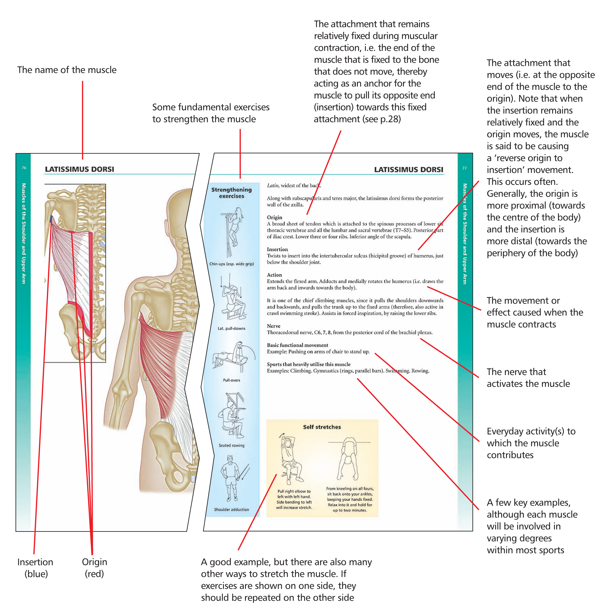
Each muscle section is colour-coded for ease of reference. Enough detail is included regarding each muscles origin, insertion and action commensurate with the requirements of the student and practitioner of bodywork, movement therapies and the movement arts. It aims to present that information accurately, in a particularly clear and user-friendly format; especially as anatomy can seem heavily laden with technical terminology. Technical terms are therefore explained in parenthesis throughout the text. The information about each muscle is presented in a uniform style throughout.
A Note About Peripheral Nerve Supply
The nervous system comprises: The central nervous system (i.e. the brain and spinal cord). the brain and spinal cord).
The peripheral nervous system (including the autonomic nervous system, i.e. all neural structures outside the brain and spinal cord). The peripheral nervous system consists of 12 pairs of cranial nerves and 31 pairs of spinal nerves (with their subsequent branches). The spinal nerves are numbered according to the level of the spinal cord from which they arise (the level is known as the spinal segment). The relevant peripheral nerve supply is listed with each muscle presented in this book, for those who need to know. However, information about the spinal segmentplexus (plexus = a network of nerves: from the Latin word meaning braid).
Therefore, such information has been derived mainly from empirical clinical observation, rather than through dissection of the body. In order to give the most accurate information possible, I have duplicated the method devised by Florence Peterson Kendall and Elizabeth Kendall McCreary (see : Muscles Testing and Function). Kendall & McCreary integrated information from six well-known anatomy reference texts; namely, those written by: Cunningham, deJong, Foerster & Bumke, Gray, Haymaker & Woodhall, and Spalteholz. Following the same procedure, and then cross-matching the results with those of Kendall & McCreary, the following system of emphasising the most important nerve roots for each muscle has been adopted in this book. Let us take the supinator muscle as our example, which is supplied by the deep radial nerve, C5, , (7). The relevant spinal segment is indicated by the letter [C] and the numbers [5, , (7)].
Bold numbers [e.g. 6] indicate that most (at least five) of the sources agree. Numbers that are not bold [e.g. 5] reflect agreement by three of four sources. Numbers not in bold and in parenthesis [e.g. (7)] reflect agreement by two sources only, or if more than two sources specifically regarded it as a very minimal supply.
If a spinal segment was mentioned by only one source, it was disregarded. Hence, bold type indicates the major innervation; not bold indicates the minor innervation; and numbers in parenthesis suggest possible or infrequent innervation.
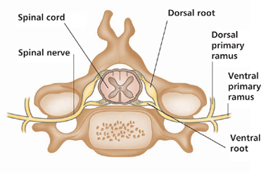
Figure 1: A spinal segment, showing the nerve roots combining to form a spinal nerve, which then divides into ventral and dorsal rami.
A spinal segment is the part of the spinal cord that gives rise to each pair of spinal nerves (a pair consists of one nerve for the left side and one for the right side of the body). Each spinal nerve contains motor and sensory fibres. Soon after the spinal nerve exits through the foramen (the opening between adjacent vertebrae), it divides into a dorsal primary ramus (directed posteriorly) and a ventral primary ramus (directed laterally or anteriorly).
Fibres from the dorsal rami innervate the skin and extensor muscles of the neck and trunk. The ventral rami supply the limbs, plus the sides and front of the trunk.
Anatomical Directions
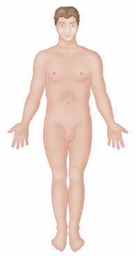
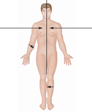
To describe the relative position of body parts and their movements, it is essential to have a universally accepted initial reference position. The standard body position known as the ). Most directional terminology used refers to the body
as if it were in the anatomical position, regardless of its actual position.
Figure 2:
Anterior.
In front of; toward or at the front of the body.
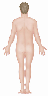
Figure 3:
Posterior.
Behind; toward or at the backside of the body.
Figure 4:
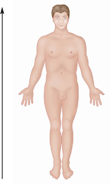
Superior.
Above; toward the head or upper part of the structure or the body.
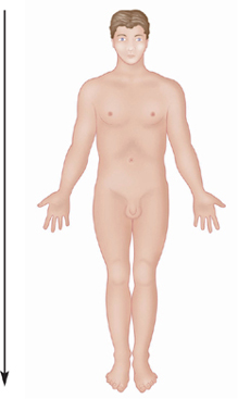
Figure 5:
Inferior.
Below; away from the head or toward the lower part of a structure or the body.


