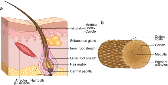
The epidermal component of the hair, called the hair shaft, is the portion of the hair that exits the scalp. The dermal components of the hair include the hair follicle (also called hair bulb or hair root) with its stem cells, blood supply, sebaceous (oil) glands, and inner and outer root sheaths (Fig. ).
Fig. 1.1
( a ) Longitudinal section of the hair depicting the epidermal and dermal components. ( b ) Cross-section of a hair shaft depicting the relationship between the three layers of the hair shaft (epidermal component)
Anatomy of the Hair: Cuticle, Cortex, and Medulla
The hair shaft is the part of the hair that is most susceptible to the effects of environmental conditions and cosmetic preparations and procedures. From the external surface inwards, the hair shaft comprises the cuticle, cortex, and medulla (Fig. ).
The Cuticle
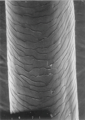
The cuticle is the outermost layer of the hair shaft and is composed of the protein keratin. It protects the underlying cortex by providing a barrier to chemicals and water []. A healthy, intact cuticle has a smooth surface and low friction in the root to tip direction and contributes to the sheen associated with healthy hair. A damaged cuticle results in hair that is frizzy, dull, and prone to breakage.
Fig. 1.2
Electron micrograph showing the overlapping, scale-like cells of the cuticle layer. (Reprinted from: Wolfram LJ. Human hair: a unique physicochemical composite. J Am Acad Dermatol. 2003;48(6 Suppl):S106-14, with permission from Elsevier)
Although the chemical composition of the cuticle is similar in all hair types, there are decreasing numbers of cuticular cell layers in Asian, Caucasian, and African hair [].
The outer aspect of the cuticle contains lipids (fatty acids, ceramides, and cholesterol) that contribute to the barrier function of the cuticle and promote the hydrophobicity and low friction of healthy hair [).
The Cortex
The majority of the mass and the tensile strength of the hair shaft can be attributed to the cortex [].
There is a strong adhesive layer between the cells of the cortex, known as the cell membrane complex (CMC ). The CMC is vulnerable to cosmetic chemical hair treatments, such as bleaching, dyeing, straightening, and perming [].
Cortical cells in human hair are divided into different regions termed orthocortex, paracortex, and mesocortex [].
Cystine and Chemical Bonds in the Cortex
There is no difference in the cystine content of keratin proteins between African hair and that of other racial groups [].
In addition to disulfide bonds, keratin proteins are also linked by weaker bonds, such as hydrogen bonds, which can be easily disrupted by water to create temporary hair styles, e.g., using rollers on wet hair to create curls (wet-setting) [].
The Medulla
The medulla forms the porous, empty center of the hair fiber (Fig. ].
In summary, a healthy hair shaft has an intact, smooth cuticle with high lipid content from root to tip and a strong cortex with intact CMCs and many disulfide bonds. These basic building blocks of healthy hair are similar in all races/ethnicities, but are vulnerable to disruption by cosmetic products and styling practices. Significant changes in the disulfide and hydrogen bonds in keratin is crucial to nearly any modification to hair, including permanent curling/straightening procedures, bleaching, wet-setting, and even daily grooming procedures such as shampooing.
Anatomy of the Hair: Dermal Structures
The Inner and Outer Root Sheaths
In the dermis, the inner root sheath (IRS) surrounds the hair shaft cuticle layer (Fig. ). External to the IRS is the outer root sheath (ORS) .
Sebaceous Glands
Conclusions from studies on racial differences in sebaceous gland size and activity are conflicting. Current opinion suggests that there are very few differences among different racial groups in this regard. However, in African hair and other curly hair types it is more difficult for sebum to make its way from the scalp down the hair shaft. Thus, curly hair types tend to be relatively dry and require regular application of cosmetic products to promote moisture retention.
Blood Supply
Studies on the racial differences of cutaneous blood supply have also shown conflicting results. However, it has been suggested that blood flow to the hair follicle in blacks is lower compared to whites, and this may contribute to the increased prevalence of scarring alopecia in black women [].
Elastic Fibers
Differences in elastic fibers among racial groups have been reported. Black patients had fewer elastic fibers anchoring their hair follicles as compared to whites []. This observation may explain black patients susceptibility to traction alopecia.

