1. Development of the Lung
The lung develops from the foregut. At the highness of the later larynx, the single tube splits into two buds for the esophagus and the lower respiratory tract, the Lungenanlage [].
The bronchial buds give rise to several generations of bronchi, starting with the main bronchi, lobar bronchi, segmental bronchi, and so on. In the human lung, approximately 16 generations are formed around the seventh week. After that, bronchioli are formed with an additional of four generations, as membranous, and three generations of terminal respiratory bronchioli. These open into alveolar ducts on which alveoli are grouped.
For the bronchial and alveolar development, the mesenchyme derived from the mesoderm is essential. Each primitive bronchus is surrounded by splanchnopleuromesoderm. Without the connection to the mesoderm, no alveoli develop [].
The different developmental stages of the lung are the embryonic stage, where the lung consists of branching tubules (gestational weeks 48). These tubules are lined by a single row of high columnar epithelium. In the pseudoglandular phase (weeks 816), the branching bronchial tree is embedded in a primitive immature mesenchyme; however, there are so many tubules that it mimics glandular structures (Figs. ]. The newborn human has approximately 50 million alveoli at birth, which represents approximately one-sixth of the number of an adult.
Fig. 1.1
Lung specimen in the early developmental tubular stage, eight week of gestation; the bronchial buds are separated by a primitive mesenchyme, only few primitive endothelial precursor cells can be identified, and capillaries have not been formed. A pulmonary artery has been cut tangentially and is seen between two bronchial buds ( right upper border to middle lower border ). H&E, bar 20 m
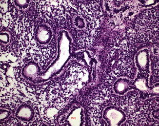
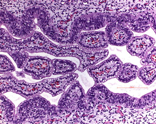
Fig. 1.2
( a , b ) Lung specimen in early developmental glandular stage, 12th gestation week; ( a ) bronchial buds are seen embedded in a primitive mesenchymal stroma, ( b ) but early glands are already formed. H&E, bar 500 and 50 m
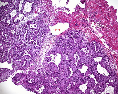
Fig. 1.3
Lung specimen in a premature child (gestation week 24); in transition from canalicular to saccular stage with primitive alveoli, which have not branched, the epithelium already shows pneumocytes in type II, and capillaries are already present; in this case the child developed bronchopulmonary dysplasia. H&E, bar 50 m
Fig. 1.4
Lung specimen at the development age of 18th gestation week; the bronchial epithelium shows nicely the clear cell pattern with apical positioned nuclei; this changes during maturation: nuclei start to move from the apical to the final basal location within the cell. The clear cell pattern results from abundant glycogen storage, which is dissolved during tissue section processing (alcohol). H&E, bar 200 m
The vascular structures arise in two different ways: the large arteries start from the sixth branchial arch and grow along the bronchial tree down to the periphery behind the ductus arteriosus. The veins develop later by sprouting from the left atrium into the mediastinum but in addition also from the sinus venosus. The veins reach the developing primitive lobules and surround them at the surface. Veins primarily form sinusoidal islands and coalesce into conducting structures following the interlobular septa [].
Bronchial arteries can be found from the ninth week of gestation. They form a plexus around the bronchi and form anastomoses with the pulmonary veins, whereas a specialized form of blood vessels, the contractile arteries, organizes the connection with the pulmonary arteries. During the saccular stage of the development, the central and peripheral vascular structures are joined. If this program is disturbed, pulmonary sequestration can result, where a part of the peripheral vascular bed is joined to a systemic artery. Also other vascular malformations such as Scimitar syndrome can be based on program failure in this period.
Lymphatic vessels are formed as a plexus in the hilar region together with the ductus thoracicus and are developed at the fifth fetal month.
Nerves are primarily formed out of ganglia of nervus vagus and truncus sympathicus/parasympathicus. An outer and inner plexus is formed around the bronchi, which is finally fused into one plexus at the site of the bronchioles. At the eight month, nerves and ganglia are mature; neurofilaments can be demonstrated. The nerves can be separated into secretory and sensory as well as motoric fibers. They are close to the bronchial muscles and also around blood vessels.
Neuroendocrine cells (NEC) can be found from the eight gestational weeks on, whereas in bronchioles and alveoli, they can be first demonstrated by neuroendocrine markers around the fifth month (chromogranin A, synaptophysin, PGP9.5). NECs are essential for the proper development and maturation of the bronchial tree.
The other mesenchymal structures, such as myoblasts and chondroblasts, develop from the coelom (splanchnopleura), which surrounds the developing bronchial tree.
The pleura also starts from the coelom (splanchnopleura), which surrounds the Lungenanlage []. From there the visceral pleura develop. From the pericardo-peritoneal channel, which is the lateral portion of the splanchnopleura, the parietal pleura arises. Primarily the parietal pleura fills both lateral thoracic cavities, since the developing bronchi occupy only small portions of the cavity. The recessus pleura pulmonalis is the only portion, which is free of lung structures.
1.1 Genetic Control of the Development
The organogenesis and maturation of the lung are under the control of genes, which are still only marginally explored. Thyroid transcription factor 1 (TTF1), hepatocyte nuclear factor (HNF3), retinoic acid receptor (RAR), Kruppel-like factor 5 (KLF5), and GATA6 all have been identified as differentiation factors for the developing lung [].
1.2 Comparison of Lung Development Across Species
Within the mammalian family, wide variations are known. In marsupials the young are born with a lung in the pseudoglandular phase; the whole lung development starts after birth. In mice, rats, and hamsters, the young are delivered with lungs in the canalicular phase, and alveoli are formed after birth. In guinea pigs and also in carnivores and sheep, the young have a fully developed lung before birth. Human beings are in between these groups: The alveolar/terminal saccular phase already starts before birth but continues after birth until the fourth to fifth year of postnatal life. After that, the lung still grows in size but the numerical structure is reached [].
References
Miura T. Modeling lung branching morphogenesis. Curr Top Dev Biol. 2008;81:291310. CrossRef PubMed