Igor Safonov Atlas of Scar Treatment and Correction 2013 10.1007/978-3-642-29196-8_1 Springer-Verlag Berlin Heidelberg 2012
1. Atrophic Scars and Stretch Marks
Igor Safonov 1
(1)
Centre of Scars Treatment and Correction, Dr. Bogomolets Institute of Dermatocosmetology, Kiev, Ukraine
Abstract
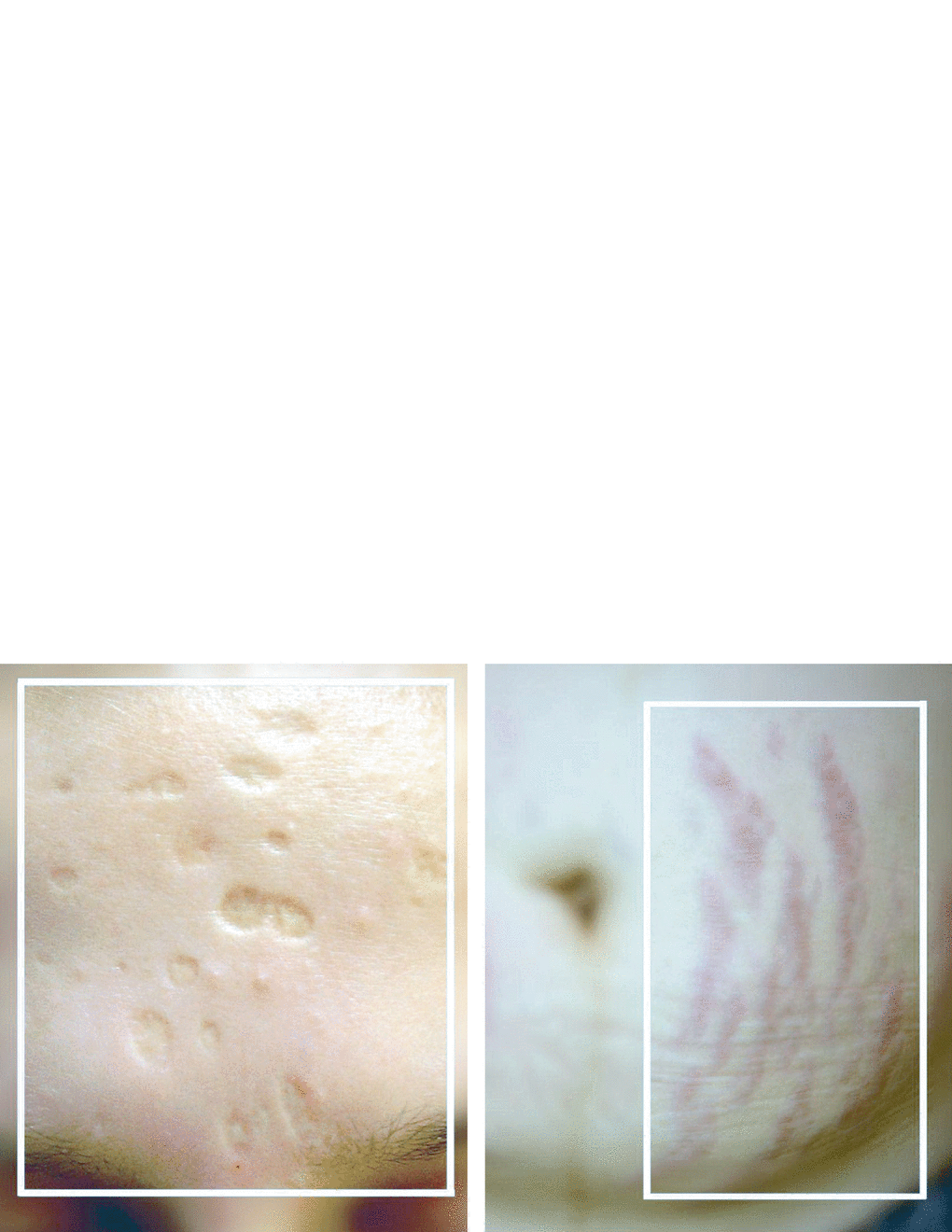
Atrophic scars are the most numerous compared to others. The common feature of these scars is their bottom, which is located below the level of surrounding tissues. The skin above atrophic scars is thin and flaccid, with cross-striation inherent in linear scars. Often, such scars are devoid of pigments and thus look white. The characteristic look of these scars is stipulated by connective tissue deficit, notably the deficit of collagen and elastin proteins that form the skin frame. This atlas places both atrophic scars and stretch marks into one section because stretch marks, by their nature, also are atrophic scars. The reason for their formation are also destroying the integrity collagen fibers and associated with this sagging skin.
Atrophic scars are the most numerous compared to others. The common feature of these scars is their bottom, which is located below the level of surrounding tissues. The skin above atrophic scars is thin and flaccid, with cross-striation inherent in linear scars. Often, such scars are devoid of pigments and thus look white. The characteristic look of these scars is stipulated by connective tissue deficit, notably the deficit of collagen and elastin proteins that form the skin frame. This atlas places both atrophic scars and stretch marks into one section because stretch marks, by their nature, also are atrophic scars. The reason for their formation are also destroying the integrity collagen fibers and associated with this sagging skin.
1.1 Atrophic Scars
1.1.1 Factors of Atrophic Scar Formation
The formation of atrophic scars may be caused by multiple reasons. The following are a number of factors provoking the formation of atrophic scars:
Loss of integrity the deep layers of the skin (dermis)
Moderate edema and inflammation
Low-intensity inflammation during phase 1 of cicatrization
Particular areas of localization
Scars resulting from animal or human bites (salivary ferments)
Postsurgery/manipulation scars (e.g., cryodestruction of hemangioma)
Acne scars
Initial debridement errors (nonfixation of wound edges)
Deficiency of oxygen, vitamin C, and other cofactors of collagen synthesis
To facilitate regular collagen synthesis, adequate delivery of oxygen, ascorbic acid and other cofactors such as zinc, iron, and copper must be ensured.
The deficiency of vitamin C in the wound area induces decreased collagen synthesis:
Procollagen triplet assembly does not take place (evidently, the helix assembly is not followed by the formation of bridges among hydroxyproline residues);
Fe2+ resorption necessary for the intracellular phase of collagen synthesis is decreased.
Intensity of Edema and Inflammation
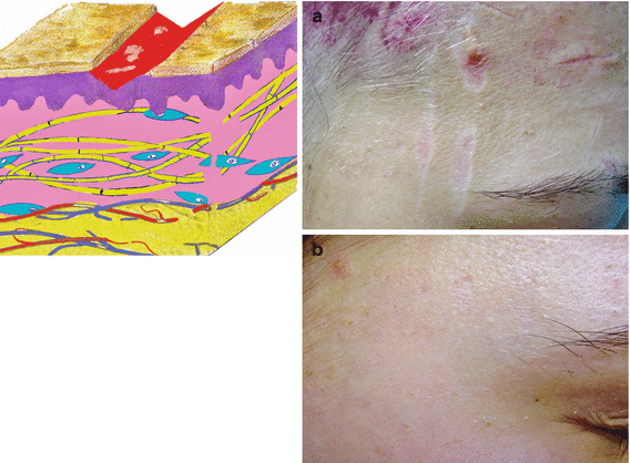
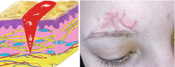
Scratches that do not reach below the papillary dermis heal without a trace, even without our interference (Fig. ). Should the injury reach the mesoderm level, a scar is inevitably formed.
Fig. 1.1
Temple scratches. ( a ) Before. ( b ) After
Atrophic scar
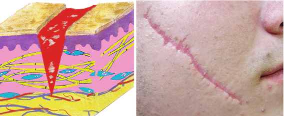
An atrophic scar is formed in the case of mild edema and sluggish inflammation (Fig. ).
Fig. 1.2
Atrophic scar of the cheek
Hypertrophic scar and keloid
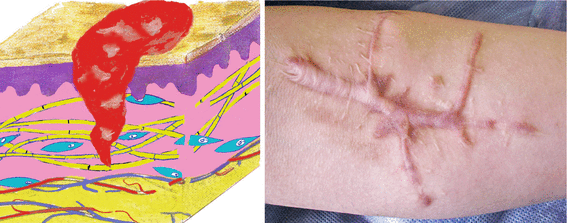
If an injury is followed by heavy bleeding, intense edema, and inflammation, such an injury is most likely to end up with a hypertrophic scar or keloid (Fig. ).
Normotrophic scar
Swelling and inflammation expressed moderately and do not go beyond the initial injury (Fig. ).
Fig. 1.4
Normotrophic scar of the forehead
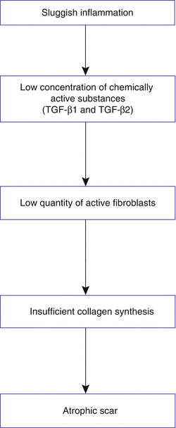
Sluggish Inflammation of Phase 1 Cicatrization
Fig. Cascade of atrophic scarring
Localization
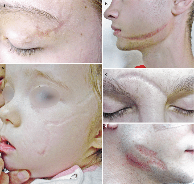
The facial muscles are mostly mimetic; therefore, atrophic scarring of the facial tissues is observed in 90% of cases after injury or surgery (Fig. )
Fig. 1.6
Postinjury facial scars. ( a ) Upper eyelid. ( b ) The area of the mandible left. () Left cheek and temple. ( d ) Forehead and interebrow area. ( e ) The area of the mandible right
Animal Bites
Atrophic scars are most likely to be formed after animal bites. Apparently, this is because the saliva rich in enzymes (including proteolytic ones) get into the wound. Bite wounds take a long time to heal, are often infected, and are hard to treat.
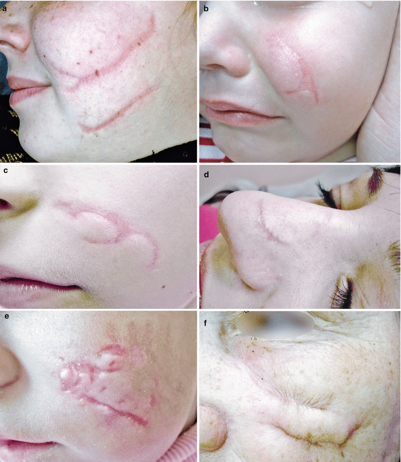
Scar edges in children and young people often dehisce because of the work of the expression muscles (Fig. ), retracts, and forms solid fibrous tissue. The treatment approach varies according to the differences mentioned.
Fig. 1.7
Animal bite. ( a f ) Left cheek bites. ( d ) Bite the nose
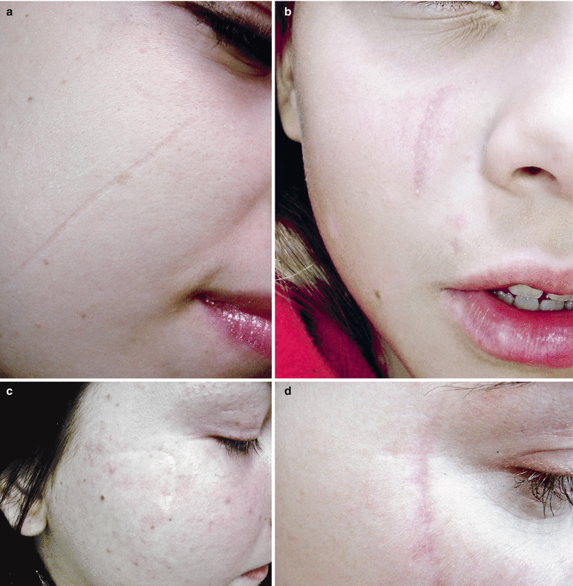
Cat-Scratch Scars
Unlike bites, cat scratches leave a small, narrow, and delicate atrophic scar (Fig. ). Such scars are hard to resurface; any resurfacing results in spreading of the scar edges, with their depth remaining unchanged because resolution of collagen tissue continuity did not take place. Consequently, there is nothing to be restored. Operative surgery is conducted within the limits of sound tissues. Thus, the postsurgery scar is wider than the one before surgery. Notwithstanding the tenuity of such scars, they tend to cause more trouble and anguish than thick scars caused by accident or surgery.
Fig. 1.8
Cat-scratch scars. ( a b ) Right cheek. ( c d ) Right zygomatic area
Medical Procedures and Operations
Figure indicates scars formed after medical procedures and operations.