• Complain

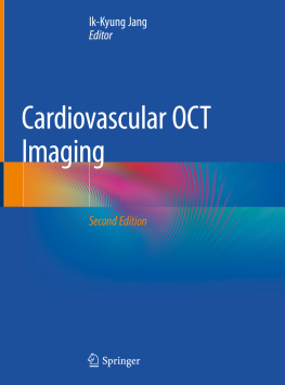
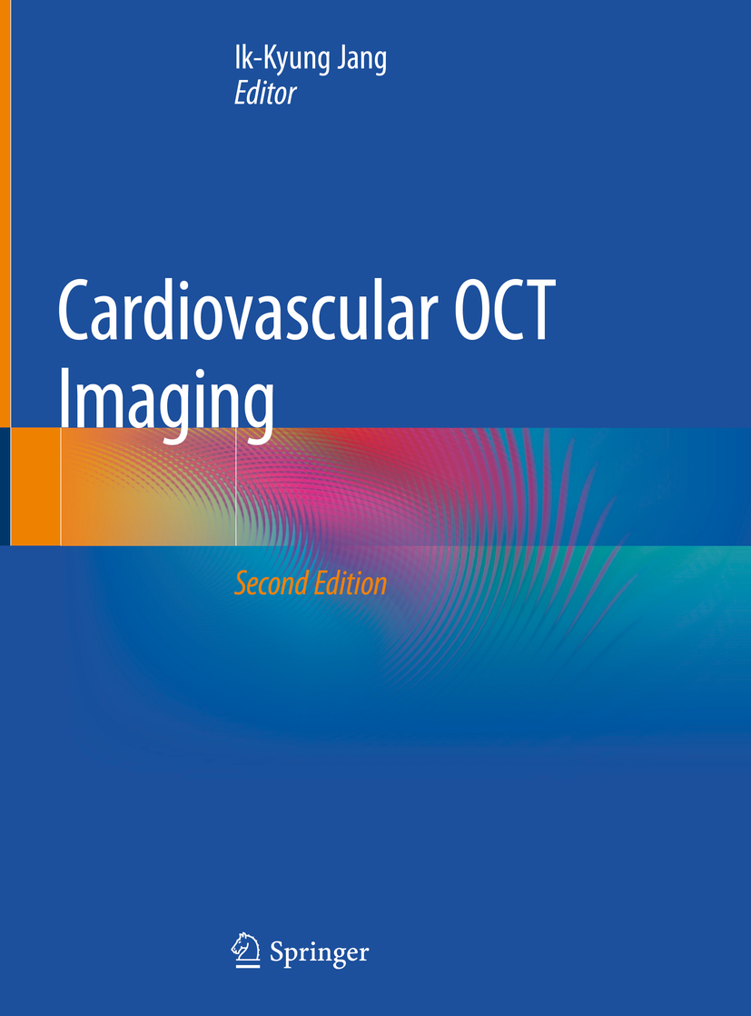
Ik-Kyung Jang - Cardiovascular OCT Imaging

Here you can read online Ik-Kyung Jang - Cardiovascular OCT Imaging full text of the book (entire story) in english for free. Download pdf and epub, get meaning, cover and reviews about this ebook. year: 2019, publisher: Springer Nature, genre: Home and family. Description of the work, (preface) as well as reviews are available. Best literature library LitArk.com created for fans of good reading and offers a wide selection of genres:

Romance novel Science fiction Adventure Detective Science History Home and family Prose Art Politics Computer Non-fiction Religion Business Children Humor

Choose a favorite category and find really read worthwhile books. Enjoy immersion in the world of imagination, feel the emotions of the characters or learn something new for yourself, make an fascinating discovery.

Ik-Kyung Jang Cardiovascular OCT Imaging
  • Book:
    Cardiovascular OCT Imaging
  • Author:
  • Publisher:
    Springer Nature
  • Genre:
  • Year:
    2019
  • Rating:
    5 / 5
  • Favourites:
    Add to favourites
  • Your mark:
    • 100
    • 1
    • 2
    • 3
    • 4
    • 5

Cardiovascular OCT Imaging: summary, description and annotation

We offer to read an annotation, description, summary or preface (depends on what the author of the book "Cardiovascular OCT Imaging" wrote himself). If you haven't found the necessary information about the book — write in the comments, we will try to find it.

This heavily revised second edition comprehensively reviews the use of optical coherence tomography (OCT) in cardiovascular practice. It provides detailed guidance on how to properly interpret OCT images and successfully utilise it in daily clinical practice Chapters cover the development and physics associated with OCT, relevant interpretation skills, OCT imaging artifacts, plaque erosion, bioabsorbable stent, the detection of vulnerable plaque, and the use of OCT imaging in unison with other modalities such as phase contrast imaging (PCI). Areas of potential future development are also covered Cardiovascular OCT Imaging enables interventional cardiologists and cardiologists to quickly become both familiar and develop a detailed understanding of this technology to improve patient care and treatment outcome. It is a valuable reference for all practising and trainee medical professionals in cardiology, and in particular those who specialize in interventional cardiology.

Ik-Kyung Jang: author's other books


Who wrote Cardiovascular OCT Imaging? Find out the surname, the name of the author of the book and a list of all author's works by series.

Cardiovascular OCT Imaging — read online for free the complete book (whole text) full work

Below is the text of the book, divided by pages. System saving the place of the last page read, allows you to conveniently read the book "Cardiovascular OCT Imaging" online for free, without having to search again every time where you left off. Put a bookmark, and you can go to the page where you finished reading at any time.

Light

Font size:

Reset

Interval:

Bookmark:

Make
Contents
Landmarks
Editor Ik-Kyung Jang Cardiovascular OCT Imaging 2nd ed 2020 Editor - photo 1
Editor
Ik-Kyung Jang
Cardiovascular OCT Imaging
2nd ed. 2020
Editor Ik-Kyung Jang Massachusetts General Hospital Harvard Medical School - photo 2
Editor
Ik-Kyung Jang
Massachusetts General Hospital Harvard Medical School, Boston, MA, USA
ISBN 978-3-030-25710-1 e-ISBN 978-3-030-25711-8
https://doi.org/10.1007/978-3-030-25711-8
Springer Nature Switzerland AG 2020
This work is subject to copyright. All rights are reserved by the Publisher, whether the whole or part of the material is concerned, specifically the rights of translation, reprinting, reuse of illustrations, recitation, broadcasting, reproduction on microfilms or in any other physical way, and transmission or information storage and retrieval, electronic adaptation, computer software, or by similar or dissimilar methodology now known or hereafter developed.
The use of general descriptive names, registered names, trademarks, service marks, etc. in this publication does not imply, even in the absence of a specific statement, that such names are exempt from the relevant protective laws and regulations and therefore free for general use.
The publisher, the authors, and the editors are safe to assume that the advice and information in this book are believed to be true and accurate at the date of publication. Neither the publisher nor the authors or the editors give a warranty, expressed or implied, with respect to the material contained herein or for any errors or omissions that may have been made. The publisher remains neutral with regard to jurisdictional claims in published maps and institutional affiliations.

This Springer imprint is published by the registered company Springer Nature Switzerland AG

The registered company address is: Gewerbestrasse 11, 6330 Cham, Switzerland

Foreword

Since the first report on OCT over two decades ago, interest in this imaging technology in medicine has exponentially increased, initially in ophthalmology and recently in cardiology. Introduction of this intravascular imaging modality in cardiology coincided with the emergence of a new concept, namely, the detection of vulnerable or high-risk plaque. As vulnerable plaque represents microstructural changes of coronary plaque including thin fibrous cap overlying lipid pool and increased lipid pool, it was thought that this high-resolution imaging modality would be ideal for detecting vulnerable plaques. Indeed, OCT has been proven to be an invaluable tool not only for detecting high-risk plaques but also for studying in vivo vascular biology, particularly in order to understand the pathophysiology of acute coronary syndromes. With a series of OCT studies, our understanding on the pathogenesis of acute coronary syndrome has made remarkable progress and confirmed many findings, which had been possible only in autopsy studies previously. Since OCT can be used in patients repeatedly, in vivo serial studies have been reported, and these studies have provided critical information on the evolution of vascular structure over time and also the vascular response to interventions such as cholesterol-lowering therapy.

In addition to the application of OCT to research, OCT has been increasingly used in interventional cardiology, primarily to optimize percutaneous coronary intervention with stenting. Despite gradual adoption of this technology in cardiac catheterization laboratories, the clinical value of OCT remains undefined. How to maximize the information provided by OCT to optimize percutaneous procedures is not widely known. This book will provide background and practical tips for interventional cardiologists.

Since his first report on in vivo OCT application in 2001, in my view, Dr. Jangs group has been the leader in OCT in cardiology. His group brought this technology from bench to bedside through series of preclinical and clinical studies. They for the first time established the OCT criteria for plaque characterization, reported differences in plaque characteristics in patients with various clinical presentations, and recently published the first in vivo demonstration of plaque erosion, one of the important underlying mechanisms for acute coronary syndromes. In addition, his group published the first paper comparing OCT and intravascular ultrasound after stenting, demonstrating the higher sensitivity of OCT in the detection of stent-related complications.

This textbook covers all the practical aspects of OCT in current research as well as clinical practice. With the rapidly growing interest in OCT, this book is extremely timely and very relevant for all cardiologists interested in vascular biology or interventional cardiology. My warmest congratulations to Dr. Jang and his group for such outstanding contributions to the field of cardiovascular medicine.

Valentin Fuster
Contents
James G. Fujimoto , Joseph Schmitt , Eric Swanson , Aaron D. Aguirre and Ik-Kyung Jang
Teruyoshi Kume , Takashi Kubo and Takashi Akasaka
Tom Adriaenssens
Jennifer E. Phipps , Taylor Hoyt , David Halaney , J. Jacob Mancuso , Sahar Elahi , Andrew Cabe , Mehmet Cilingiroglu , Thomas E. Milner and Marc D. Feldman
Francesco Fracassi and Giampaolo Niccoli
Vikas Thondapu , Peter Libby and Ik-Kyung Jang
Ashkan Parsa and Jacqueline Saw
Teruyoshi Kume and Shiro Uemura
Taishi Yonetsu
Tom Adriaenssens
Alessio Mattesini , Antonio Martellini , Luigi Tassetti and Carlo Di Mario
Rocco Vergallo and Ik-Kyung Jang
Kensuke Nishimiya and Guillermo Tearney
Martin Villiger , Jian Ren , Nstor Uribe-Patarroyo and Brett E. Bouma
Springer Nature Switzerland AG 2020
I.-K. Jang (ed.) Cardiovascular OCT Imaging https://doi.org/10.1007/978-3-030-25711-8_1
1. The Development of Optical Coherence Tomography
James G. Fujimoto
(1)
Department of Electrical Engineering and Computer Science, Research Laboratory of Electronics, Massachusetts Institute of Technology, Cambridge, MA, USA
(2)
Health Technologies, Apple, Inc., Sunnyvale, CA, USA
(3)
Research Laboratory of Electronics, Massachusetts Institute of Technology, Cambridge, MA, USA
(4)
Division of Cardiology, Massachusetts General Hospital, Harvard Medical School, Boston, MA, USA
James G. Fujimoto (Corresponding author)
Email:
Joseph Schmitt
Eric Swanson
Email:
Aaron D. Aguirre
Email:
Ik-Kyung Jang
Email:
Keywords
Optical coherence tomography OCT Optical biopsy Intravascular OCT Swept source OCT Fourier domain OCT Optical frequency domain imaging OFDI Fiber optic catheter
1.1 Introduction

Optical coherence tomography (OCT) has become a major optical imaging modality in biomedical optics and medicine. OCT performs high resolution, cross-sectional and three dimensional volumetric and functional imaging of the internal microstructure in biological tissues by measuring backscattered light []. Tissue pathology can be imaged in situ and in real time with resolutions of 115 m, one to two orders of magnitude finer than conventional ultrasound. Imaging can be performed using small fiber-optic probes including catheters and endoscopes. The unique features of OCT make it a powerful imaging modality with applications spanning many clinical specialties as well as fundamental scientific and biological research and many non-medical applications.

Next page
Light

Font size:

Reset

Interval:

Bookmark:

Make

Similar books «Cardiovascular OCT Imaging»

Look at similar books to Cardiovascular OCT Imaging. We have selected literature similar in name and meaning in the hope of providing readers with more options to find new, interesting, not yet read works.


Reviews about «Cardiovascular OCT Imaging»

Discussion, reviews of the book Cardiovascular OCT Imaging and just readers' own opinions. Leave your comments, write what you think about the work, its meaning or the main characters. Specify what exactly you liked and what you didn't like, and why you think so.