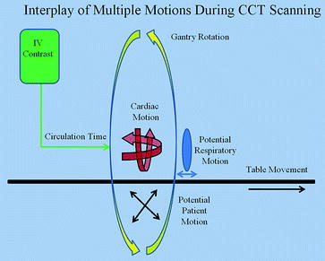
Meticulous attention to the methodology of image acquisition and reconstruction is essential to performance and analysis of coronary artery CT angiography. High-quality images must be captured during the most quiescent phase of coronary artery motion while minimizing patient exposure to radiation and intravenous contrast agents. Successful imaging requires an understanding of the temporal interplay of multiple motions including the complexities of cardiac contraction, potential respiratory motion, potential patient movement, table motion, gantry rotation, and timing and movement of the bolus of intravenous contrast through the structures of interest (Fig.). Additional factors relate to the use of x-ray energy to capture adequate images balanced by the limitation of radiation to the lowest level possible.
Fig. 1.1
The multiple motions during cardiac CT acquisition. Red signifies the cardiac motion. Yellow represents the gantry movement (rotation of the x-ray tube and detectors around the patient). Black represents the table motion and blue represents potential respiratory motion. An additional consideration is the time from the vein injection to the arrival of contrast in the coronary circulation (green)
1.1 Concepts for Image Acquisition
There are many interdependent factors that affect spatial and temporal resolution. The goal of image acquisition is to visualize the target structures in their entirety while limiting the field of view (FOV) to the region of clinical interest. For a given voxel matrix, smaller FOVs yield smaller voxels and greater spatial resolution. Voxel attenuation is measured (on a scale) with the attenuation values of different substances represented by different Hounsfield units (HU) values []. Representative HU values include the following: air 1000, fat 50 to 100, water 0, muscle 1040, contrast 300500, and calcium 1301500.
Conceptually, all multidetector computed tomography (MDCT) systems work using similar principles, but vary in regard to specific components and features. A MDCT system has an x-ray tube that is capable of extremely rapid rotation and a collimated array of detectors. The x-ray tube provides radiation energy adjustable through tube current (mAs) and tube voltage (kVP). Multiple rows of detectors are arranged in a variety of arrays with the goal of covering a specified volume during each gantry rotation. Advances in detector number and arrangement have lead to increases in the volume of coverage per rotation, with the ultimate goal being imaging the entire heart within one cardiac cycle.
The relationship between table movement, gantry rotation speed, and beam collimation defines the volume coverage per rotation as well as the degree of overlap between rotations. The concept of pitch quantifies this relationship. Pitch relates to coverage obtained by the x-ray beam (through beam width and table movement) during one rotation of the gantry and defines the amount of overlap of acquired data. With a pitch value of less than 1, there is overlap between volumes of coverage. Overlapping images allow for oversampling, permitting multisector image reconstruction, at the expense of greater radiation exposure. The definition of pitch has evolved with newer scanner technologies, and various equations have been proposed depending on the specific type of scanner []. Newer systems have thinner slice thickness and collimation, allowing for an even lower pitch resulting in more images, thinner reconstruction intervals, and better visualization of the coronary anatomy.
ECG triggering is essential to minimize the effects of cardiac motion on image acquisition. Factors related to motion in the X, Y, and Z directions include coronary artery and cardiac motion during the cardiac cycle related to heart rate and rhythm. In MDCT, images are usually acquired with retrospective ECG gating. With this approach, images are acquired throughout the cardiac cycle, and therefore can be reconstructed at specific points in the cardiac cycle []. The disadvantage relates to the limited dataset obtained. If the images obtained demonstrate significant motion artifact, there are no other images to reconstruct. Given the variability of heart rate with arrhythmias, prospective gating can be problematic with significant ectopy or atrial fibrillation.
Breath hold is essential to limit motion of structures due to respiration during image acquisition. Breath hold times have decreased significantly with advances in technology and are in the range of 610 second. There is some controversy as to the optimal phase of respiration for breath hold. Regardless of the phase chosen in an individual lab, it is important to practice breath hold commands and exercises prior to the scan. As an end-inspiratory breath hold will place structures more caudally than an end-expiratory breath hold, consistent breath hold instructions need to be given for preview images and actual scans.
1.2 Preview Scans
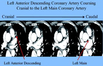
Cardiovascular CT is performed using planar scout images, a non-contrast calcium scan, a timing scan for assessment of the circulation time, and a contrast scan. Planar scout images are obtained in order to define the most cranial and caudal scanning levels (z-axis) of the structures of interest. The scout images are obtained in antero-posterior and lateral views and aligned to the patient by a laser system. The scan volume is selected with the structures of interest placed within the center of the scanning volume. Important landmarks can be identified including the left atrial appendage, which is usually the most cranial structure of the heart, and the ventricular apex, which is the most caudal structure. Although the carina serves as a marker to localize the most cranial aspect of the heart, the distance from carina to left main coronary artery is extremely variable [ ). For coronary artery imaging, scanning 10 mm cranial to the left main coronary artery and 10 mm caudal to the apex is subsequently performed with CT angiography. In patients with coronary artery bypass grafts, the starting point is the top of the aortic arch or 10 mm higher than the surgical metal clips. The mid-level of the right pulmonary artery can also be used as the beginning of the scan level, if it can be defined in preview images.
After the scout images, a calcium scan is performed. This is a high-resolution non-contrast cardiac-gated study, which provides important prognostic information regarding future cardiovascular risk. For the calcium scan, the 2-D axial images are analyzed with the identification of calcium using either manual or automated methods with quantification of calcium score based on identification of HU units with an attenuation of at least 130 HU in the areas of identified calcium. There are two major methods of quantifying coronary artery calcium: the Agatston score and the volumetric analysis. The Agatston score is based on the summation of plaque area times a coefficient based on the peak HU units in the plaque for all identified plaques [].

