Marc Dewey Cardiac CT 10.1007/978-3-642-14022-8_1 Springer-Verlag Berlin Heidelberg 2011
1. Introduction
Abstract
The advent of multislice computed tomography was a quantum leap for CT technology. When this technical innovation was first introduced, the radiological community was faced with the task of putting its advantages to use for diagnostic patient management and optimizing its clinical applications. One of the major clinical challenges was to develop this new tool for noninvasive cardiac imaging applications ranging from coronary angiography, to ventricular function analysis, to cardiac valve evaluation.
The advent of multislice computed tomography was a quantum leap for CT technology. When this technical innovation was first introduced, the radiological community was faced with the task of putting its advantages to use for diagnostic patient management and optimizing its clinical applications. One of the major clinical challenges was to develop this new tool for noninvasive cardiac imaging applications ranging from coronary angiography, to ventricular function analysis, to cardiac valve evaluation.
Marc Dewey and the authors of the book have closely followed the development of this new generation of CT scanners in the clinical setting, in scientific studies, and in experimental investigations. The team of authors has gained a wealth of experience spanning CT from 16-row technology to the most recent generation of 320-row CT scanners. In their scientific investigations, the authors have always placed great emphasis on a critical appraisal of this emerging imaging modality in comparison to well-established diagnostic tests such as coronary angiography, magnetic resonance imaging, and echocardiography, also including the socioeconomic perspective. The close cooperation with the Departments of Cardiology and Cardiac Surgery of the Charit Universittsmedizin Berlin was pivotal for obtaining valid results in both clinical examinations and scientific studies and also led to many improvements of the diagnostic workflow.
This book focuses on how to integrate cardiac CT into routine practice. Readers will learn how to perform noninvasive imaging of the heart using CT and how to interpret the images. A clear overview of the essentials is given, and numerous clinical cardiac CT cases are presented for illustration.
All steps involved in cardiac CT examination are described in detail, including patient preparation, the actual examination, and analysis and interpretation of the findings. Cardiac CT is based on an earlier book Coronary CT Angiography but also updates all chapters of this book and includes completely new chapters on radiation exposure, clinical practice, coronary artery stents, bypass grafts, coronary anomalies, and congenital heart disease. Separate chapters discuss upcoming clinical applications of cardiac CT plaque imaging and assessment of cardiac functions and valves. Another asset of the book in terms of practical clinical application is that the authors present and discuss the specific features of the CT scanners from all four major vendors as they relate to cardiac imaging. In a final chapter, an outlook is given on conceivable future technical and clinical developments.
I congratulate the team of authors on an excellent book that focuses on the practical clinical aspects of cardiac CT and offers its readers an easy to follow introduction to this promising new diagnostic tool. However, the book also provides useful tips and tricks for those already familiar with this imaging modality, which will help them further improve their diagnostic strategy for the benefit of their patients.
Marc Dewey Cardiac CT 10.1007/978-3-642-14022-8_2 Springer-Verlag Berlin Heidelberg 2011
2. Technical and Personnel Requirements
2.1 Technical Requirements
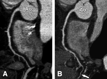
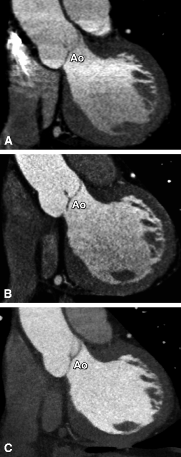
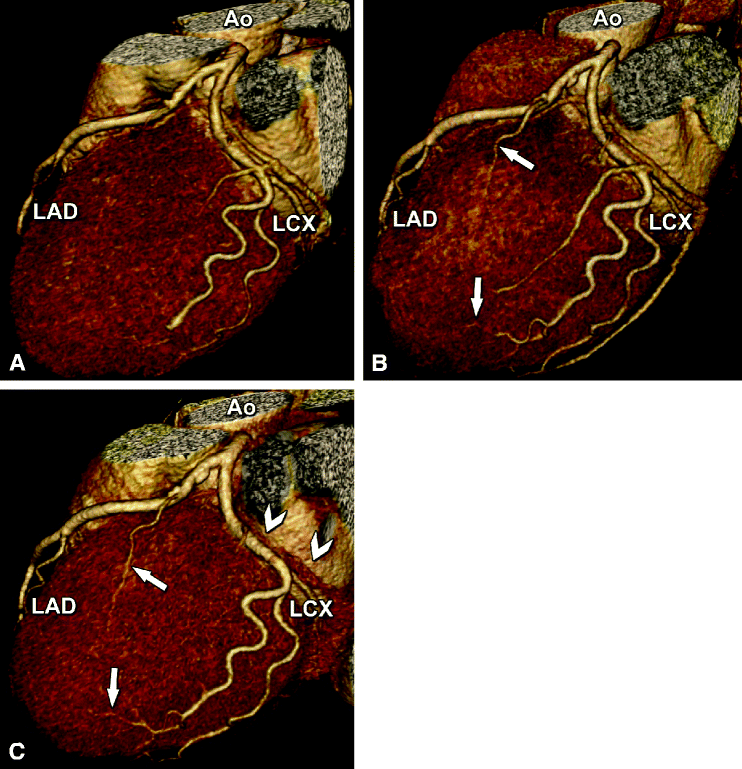
Noninvasive coronary angiography is an ascending clinical application that requires very high spatial and temporal resolution. Thus, CT scanners with multiple detector rows (multislice CT [MSCT]), short gantry rotation times, and thin-slice collimation are essential for establishing a successful cardiac CT imaging center. Because 64-row CT is superior to 16-row CT in terms of image quality and diagnostic accuracy, we believe that (at least) 64-row technology should be used for noninvasive coronary angiography ( List 2.1 ). CT with 64-row technology not only increases the quality of the images ( Figs. ). The faster gantry rotation speed of recent CT scanners ( List 2.1 ) improves temporal resolution and dramatically reduces the likelihood of relevant motion artifacts.
Fig. 2.1
Comparison of 16-row (Panel A) and 64-row CT coronary angiography ( Panel B ) of the right coronary artery (curved multiplanar reformation) in a 61-year-old male patient. 64-row CT shows longer vessel segments, especially in the periphery (arrow). This enhanced performance can be explained by fewer motion artifacts (due to breathing, extrasystoles, or variations in the length of the cardiac cycle) and the better contrast between arteries and veins resulting from the faster scan and consequently better depiction of the arterial phase. The improved depiction of the arterial phase using 64-row CT is also demonstrated in Fig. . Panel B also illustrates the slightly higher image noise with 64-row CT, which can be compensated for by the better depiction of the arterial phase and the higher intravascular density. Ao aorta
Fig. 2.2
The improved depiction of the arterial phase using 64-row CT ( Panel B ) and even further using 320-row CT ( Panel C ) when compared with 16-row CT ( Panel A ) is illustrated by a double-oblique coronal slice along the left ventricular outflow tract, with the aortic valve nicely depicted (Ao). In the craniocaudal direction, the density in the aorta and left ventricle shows less variation and decline when 64 simultaneous detector rows are used ( Panel B ) and almost no difference with 320-row CT acquired during a single heartbeat. Use of 64- and 320-row CT thus improves image quality and facilitates the application of automatic coronary vessel and cardiac function analysis tools
Fig. 2.3
Example illustrating the improved depiction of distal coronary artery branches using 64-row ( Panel B ) and 320-row CT ( Panel C ) in a 58-year-old female patient. Three-dimensional volume-rendered reconstructions of the left coronary artery with the left anterior descending (LAD) and left circumflex coronary artery (LCX) examined using 16-row ( Panel A ), 64-row ( Panel B ), and 320-row CT coronary angiography ( Panel C ). Note the improved depiction of smaller side branches with the 64-row ( arrows in Panel B ) and 320-row technology ( arrows in Panel C ) when compared with the same segments in 16-row CT ( Panel A ). Also, there is best depiction of the arterial phase (with less venous overlap, arrowheads in Panel C ) using 320-row CT. Single-beat imaging using 320-row CT or second-generation dual-source CT with a fast prospective spiral also greatly reduces radiation exposure (). Ao aorta

