• Complain

Franco Rongioletti - Rare Malignant Skin Tumors

Here you can read online Franco Rongioletti - Rare Malignant Skin Tumors full text of the book (entire story) in english for free. Download pdf and epub, get meaning, cover and reviews about this ebook. year: 2015, publisher: Springer, genre: Home and family. Description of the work, (preface) as well as reviews are available. Best literature library LitArk.com created for fans of good reading and offers a wide selection of genres:

Romance novel Science fiction Adventure Detective Science History Home and family Prose Art Politics Computer Non-fiction Religion Business Children Humor

Choose a favorite category and find really read worthwhile books. Enjoy immersion in the world of imagination, feel the emotions of the characters or learn something new for yourself, make an fascinating discovery.

Franco Rongioletti Rare Malignant Skin Tumors

Rare Malignant Skin Tumors: summary, description and annotation

We offer to read an annotation, description, summary or preface (depends on what the author of the book "Rare Malignant Skin Tumors" wrote himself). If you haven't found the necessary information about the book — write in the comments, we will try to find it.

Franco Rongioletti: author's other books


Who wrote Rare Malignant Skin Tumors? Find out the surname, the name of the author of the book and a list of all author's works by series.

Rare Malignant Skin Tumors — read online for free the complete book (whole text) full work

Below is the text of the book, divided by pages. System saving the place of the last page read, allows you to conveniently read the book "Rare Malignant Skin Tumors" online for free, without having to search again every time where you left off. Put a bookmark, and you can go to the page where you finished reading at any time.

Light

Font size:

Reset

Interval:

Bookmark:

Make
Part I
Tumours of the Epidermis
Springer Science+Business Media New York 2015
Franco Rongioletti , Irina Margaritescu and Bruce R Smoller Rare Malignant Skin Tumors 10.1007/978-1-4939-2023-5_1
1. Adenoid (Acantholytic) Squamous Cell Carcinoma
Valentina Caputo 1
(1)
Department of Surgical Pathology, Fatebenefratelli, Via M. Melloni 52, Milan, 20219, Italy
(2)
Section of Dermatology, Department of Health Sciences and Division of Pathology, University of Genova, Genova, Italy
Valentina Caputo (Corresponding author)
Email:
Franco Rongioletti
Email:
Email:
Keywords
Adenoid squamous cell carcinoma Pseudoglandular squamous cell carcinoma Epithelioma spinocellular segregans
Introduction
Cutaneous acantholytic squamous cell carcinoma (ASCC) was initially described in 1947 by Lever as a tumor composed of both solid and gland-like epithelial nests and was called adenoacanthoma of the sweat glands. He later revised his classification, and actually, ASCC is considered an uncommon, distinctive variant of squamous cell carcinoma, characterized by a typical SCC pattern in combination with acantholytic pseudoglandular space formation. Although ASCC is considered as a more aggressive variant of SCC, its prognosis seems mostly to be more related to the characteristics of the host and the grading of tumor rather than the histopathological features.
Clinical Features
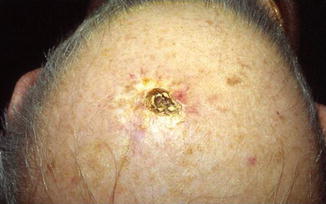
ASCC generally presents as a reddish, scaling, often cup-shaped, and ulcerated lesion on sun-exposed areas of elderly patients, predominantly on the head (Fig. ) and neck, although other sites such as the vulva, penis, breast, foot, leg, and arm can be involved. It is more common in males than females, with a mean age at diagnosis of 70 years.
Fig 11 A keratotic nodule with erythematous halo on the scalp of an old - photo 1
Fig. 1.1
A keratotic nodule with erythematous halo on the scalp of an old patient
Pathology
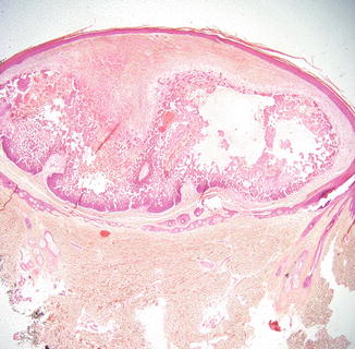
Histologically, the tumor is composed of cords and nests of atypical keratinocytes with an infiltrative pattern of growth. Connection to the overlying epidermis, which may show hyperkeratosis and parakeratosis, is present in most cases, even if sometimes it may be only focal or absent (Fig. ). ASCC stains positively for immunohistochemical markers towards pankeratin and epithelial membrane antigen (EMA), but immunostains for carcinoembryonic antigen (CEA) give invariably negative results. In the literature, a decreased expression of intercellular adhesion proteins, such as of Dsg3, E-cadherin, and syndecan-1, has been described in ASCC compared to conventional SCC, and it has been suggested that this may contribute to the development of acantholysis and of the controversially more aggressive biological behavior.
Fig 12 A squamous tumor extending into the dermis with pseudoglandular spaces - photo 2
Fig. 1.2
A squamous tumor extending into the dermis with pseudoglandular spaces and necrotic areas
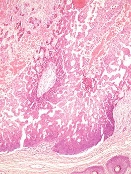
Fig 13 Pseudovascular or pseudoglandular appearance due to tumor cell - photo 3
Fig. 1.3
Pseudovascular or pseudoglandular appearance due to tumor cell discohesiveness and necrosis
Fig 14 Lining is composed of squamous epithelium and spaces with necrotic - photo 4
Fig. 1.4
Lining is composed of squamous epithelium and spaces with necrotic debris, keratin, and keratinous cyst
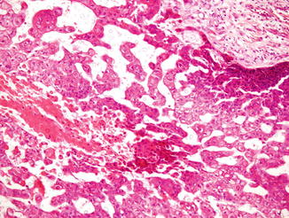
Fig 15 The epithelial cells are atypical with bright eosinophilic focally - photo 5
Fig. 1.5
The epithelial cells are atypical with bright eosinophilic, focally glassy cytoplasm arranged in loose cohesive clusters representing pseudoacantholysis
Acantholytic cells may appear extremely bizarre, large, with glassy eosinophilic cytoplasm. Nuclei may vary from atypical to pleomorphic and may be single or multiple, and mitotic figures are variably present.
Differential Diagnosis
Differential diagnosis is only histological and is directed mainly towards gland-forming malignancies, as eccrine adenocarcinoma, adenosquamous carcinoma, and metastatic adenocarcinoma. Another important differential diagnosis concerns epithelioid angiosarcoma. In eccrine adenocarcinoma as in adenosquamous carcinoma, the glandular spaces are lined with periodic acidSchiff (PAS)-positive cells, whereas in ASCC, the cells are PAS negative. Moreover, ASCC shows no mucin secretion, and pseudoglandular spaces are due to acantholysis, not to real glandular differentiation, and tumor cells in ASCC lack the immunohistochemical positivity to CEA, S100 protein, and keratin 7 that can be seen in glandular malignancies. In epithelioid angiosarcoma, the vascular spaces contain red blood cells and not acantholytic keratinocytes, although cases of ASCC containing red blood cells within the pseudoglandular spaces have been notoriously described. In these cases, immunohistochemical stains are required. Angiosarcomas are typically positive for vimentin and CD-34, whereas ASCC shows positivity for cytokeratin and EMA.
Prognosis
It is difficult to assess the real biological behavior and metastatic potential of ASCC, because of the lack of adequate data regarding tumor size and site and circumstances of the patients in several of the largest published series. In the literature, the recurrence and metastasis rates reported are variable and range from 6 % to 10 % and from 2 % to 43 %, but, according to a recent analysis, the aggressive biological behavior of ASCC seems to correlate more with factors such as tumor size greater than 1,5 cm, recurrence, site of involvement, infiltration depth >3 mm, immunosuppression, and radiation therapy rather than with histopathological features. Moreover, recurrent tumors and metastases do not always reproduce the adenoid pattern of origin.
Treatment
The treatment and follow-up of the patients should be similar to that for conventional SCC. Early wide surgical excision of the tumor has to be considered the treatment of choice.
Bibliography
Cassarino DS, Derienzo DP, Barr RJ. Cutaneous squamous cell carcinoma: a comprehensive clinicopathologic classification. Part one. J Cutan Pathol. 2006;33:191206.
Garcia C, Crowson AN. Acantholytic squamous cell carcinoma: is it really a more-aggressive tumor? Dermatol Surg. 2011;37:3536. PubMed CrossRef
Griffin JR, Wriston CC, Peters MS, Lehman JS. Decreased expression of intercellular adhesion molecules in acantholytic squamous cell carcinoma compared with invasive well-differentiated squamous cell carcinoma of the skin. Am J Clin Pathol. 2013;139:4427. PubMed CrossRef
Lever WF. Adenoacanthoma of sweat glands: carcinoma of sweat glands with glandular and epidermal elements-report of four cases. Arch Dermatol Syphilol. 1947;56:15771. CrossRef
Nagor E, Sanchez-Motilla JM, Perez-Valles A, Martinez-Lahuerta C, Alegre V, Aliaga A. Pseudovascular squamous cell carcinoma of the skin. Clin Exp Dermatol. 2000;25:2068. CrossRef
Next page
Light

Font size:

Reset

Interval:

Bookmark:

Make

Similar books «Rare Malignant Skin Tumors»

Look at similar books to Rare Malignant Skin Tumors. We have selected literature similar in name and meaning in the hope of providing readers with more options to find new, interesting, not yet read works.


Reviews about «Rare Malignant Skin Tumors»

Discussion, reviews of the book Rare Malignant Skin Tumors and just readers' own opinions. Leave your comments, write what you think about the work, its meaning or the main characters. Specify what exactly you liked and what you didn't like, and why you think so.