• Complain

Emilio Quaia - Radiological Imaging of the Kidney

Here you can read online Emilio Quaia - Radiological Imaging of the Kidney full text of the book (entire story) in english for free. Download pdf and epub, get meaning, cover and reviews about this ebook. City: Berlin;Heidelberg, year: 2011, publisher: Springer, genre: Home and family. Description of the work, (preface) as well as reviews are available. Best literature library LitArk.com created for fans of good reading and offers a wide selection of genres:

Romance novel Science fiction Adventure Detective Science History Home and family Prose Art Politics Computer Non-fiction Religion Business Children Humor

Choose a favorite category and find really read worthwhile books. Enjoy immersion in the world of imagination, feel the emotions of the characters or learn something new for yourself, make an fascinating discovery.

Emilio Quaia Radiological Imaging of the Kidney
  • Book:
    Radiological Imaging of the Kidney
  • Author:
  • Publisher:
    Springer
  • Genre:
  • Year:
    2011
  • City:
    Berlin;Heidelberg
  • Rating:
    4 / 5
  • Favourites:
    Add to favourites
  • Your mark:
    • 80
    • 1
    • 2
    • 3
    • 4
    • 5

Radiological Imaging of the Kidney: summary, description and annotation

We offer to read an annotation, description, summary or preface (depends on what the author of the book "Radiological Imaging of the Kidney" wrote himself). If you haven't found the necessary information about the book — write in the comments, we will try to find it.

Emilio Quaia: author's other books


Who wrote Radiological Imaging of the Kidney? Find out the surname, the name of the author of the book and a list of all author's works by series.

Radiological Imaging of the Kidney — read online for free the complete book (whole text) full work

Below is the text of the book, divided by pages. System saving the place of the last page read, allows you to conveniently read the book "Radiological Imaging of the Kidney" online for free, without having to search again every time where you left off. Put a bookmark, and you can go to the page where you finished reading at any time.

Light

Font size:

Reset

Interval:

Bookmark:

Make
Part 1
Embryology and Anatomy
Emilio Quaia (ed.) Medical Radiology Radiological Imaging of the Kidney 10.1007/978-3-540-87597-0_1 Springer Berlin Heidelberg 2010
1. Embryology of the Kidney
Marina Zweyer 1
(1)
Clinical Department of Biomedicine, University of Trieste, via Manzoni 16, 34138 Trieste, Italy
Marina Zweyer
Email:
1.1
1.2
1.3
1.3.1
1.3.2
1.4
1.5
1.6
1.7
1.8
1.8.1
1.8.2
1.9
1.9.1
1.9.2
1.9.3
1.9.4
1.9.5
1.9.6
1.9.7
1.10
Abstract
During the third week of pregnancy, the process of gastrulation results in the formation of trilaminar embryo in humans. This is the beginning of morphogenesis (development of body form). Three germ layers, ectoderm, mesoderm, and endoderm, are present. A specific part of mesoderm, the intermediate mesoderm, gives rise to urogenital system gradually. It starts to develop from nephrogenic cord, which divides in nephrotomes. They give rise to pronephroi and subsequently mesonephroi, the transitory kidneys. Finally, metanephroi appear as the permanent organs, from two different structures: the metanephrogenic blastema (mesenchimal component) that leads to nephrons, and ureteric bud (epithelial component) that gives rise to collecting tubules, calices, renal pelvis, and ureter. Following developmental steps depend on inductive signaling between metanephrogenic blastema and ureteric bud.
Many genes, regulating proteins and pathways are involved in the physiological organogenesis. Cell proliferation and apoptosis keep the balance of the growth. Defects in these molecular and morphogenetic mechanisms may cause various congenital abnormalities of the kidney and urinary tract (CAKUT), which represent a family of diseases with a diverse anatomical spectrum.
1.1 Introduction
The morphological and functional complexity of the mammalian kidney arises from the highly coordinate series of events that lead to its development.
The study of structural and molecular aspects of kidney formation has been carried out for a great part in humans. Nevertheless, the involvement of specific genes, proteins, and pathways in the patterning of kidney has been disclosed mainly by genetic ablation in experiments in mouse. The definition of early events that lead to kidney primordium has also been realized by investigating the processes in nonmammalian vertebrate species, like fish, frog, and chick embryos. These integrated studies have been possible for the conservation of many genes in the different vertebrates and for the common origin of their embryonic kidney.
For many decades, the researchers focused their attention on morphological aspects of the development. The first studies about cellular basis of embryogenesis have been carried out by Grobstein (), which have introduced the modern kidney embryology, using a mammalian in vitro organ culture system.
Recently, several authors excellently reviewed all the studies on the molecular basis of renal embryogenesis (Lehner and Dressler ).
Many investigations have, moreover, been focused on the congenital abnormalities of the kidney and urinary tract (CAKUT), and their origin from morphogenetic defects (Kuwajama et al. ; Schedl 2007).
Here we describe the basic structural events of kidney embryogenesis in the context of urinary system and provide a synthesis of the molecular basis of morphogenesis. Congenital anomalies of the kidney and urinary tract caused by an uncorrected development are finally presented.
1.2 Development of Urogenital System
During the third week of the development, the embryo is composed of three germ layers, ectoderm, mesoderm, and endoderm. Each of them gives rise to specific tissue and organs. Ectoderm is the source of the epidermis, the nervous system, and several other structures. Endoderm principally gives rise to epithelial component of the respiratory and digestive tracts, including associated glands. Mesoderm is the source of connective tissues, smooth and striated muscles, cardiovascular system and blood, bone marrow, skeleton, and the reproductive and excretory systems. Mesoderm differentiates in three parts on each side of the embryo: beginning from the embryonic axis, the paraxial mesoderm, which gives rise to the somites, the intermediate mesoderm, and lateral mesoderm. In particular, the intermediate mesoderm plays a fundamental role in the development of urogenital apparatus.
The urogenital apparatus is functionally divided into the urinary (excretory) system and the genital (reproductive) system. During development, these two systems are tightly associated. Moreover, they are anatomically connected in adult males because both urine and semen pass through the urethra. In adult females, the urethra and vagina are separate, but open into a common cavity, the vestibule of the vagina (Moore ). The suprarenal glands , originated from lateral mesoderm and neural crests, are topographically associated to the superior poles of the kidneys, and its congenital hyperplasia causes anomalies both in female and male external genitalia. Intermediate mesoderm extends along the dorsal wall of the embryo.
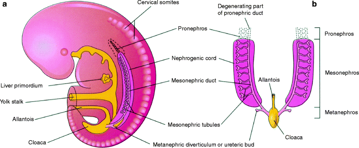
During folding of the embryo in the horizontal plane, this mesoderm component is carried ventrally. A longitudinal elevation of the mesoderm called urogenital ridge forms on each side of the dorsal aorta. It gives rise to the urinary and genital systems. Nephrogenic ridge or cord (Fig. b. The urinary system begins to develop before the genital system.
Fig 11 Diagrammatic sketches illustrating the three sets of excretory systems - photo 1
Fig. 1.1
Diagrammatic sketches illustrating the three sets of excretory systems in an embryo during the fifth week. ( a ) Lateral view. ( b ) Ventral view. The mesonephric tubules have been pulled laterally; their normal position is shown in ( a )
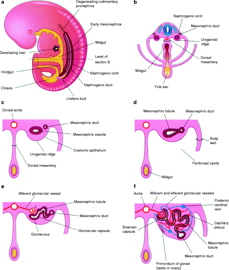
Fig 12 a Sketch of a lateral view of a 5-week embryo showing the extent - photo 2
Fig. 1.2
( a ) Sketch of a lateral view of a 5-week embryo showing the extent of the mesonephros and the primordium of the metanephros or permanent kidney. ( b ) Transverse section of the embryo showing the nephrogenic cords from which the mesonephric tubules develop. Observe the position of the urogenital ridges and nephrogenic cords. ( c f ) Sketches of transverse sections showing successive stages in the development of a mesonephric tubule between the fifth and eleventh weeks. Note that the mesenchymal cell cluster in the nephrogenic cord develops a lumen, thereby forming a mesonephric vesicle. The vesicle soon becomes an S-shaped mesonephric tubule and extends laterally to join the pronephric duct, now renamed the mesonephric duct. The expanded medial end of the mesonephric tubule is invaginated by blood vessels to form a glomerular capsule (Bowman capsule). The cluster of capillaries projecting into this capsule is the glomerulus
1.3 Development of Urinary System
The urinary tract is composed of:
  • The kidneys , the main filtrating organs in the mammalian organisms; the morphofunctional unit of the kidney is the nephron. It provides for blood filtration, reabsorption of water, salt, and other substances necessary for the organism. The nephrons modulate blood pressure and pH.
Next page
Light

Font size:

Reset

Interval:

Bookmark:

Make

Similar books «Radiological Imaging of the Kidney»

Look at similar books to Radiological Imaging of the Kidney. We have selected literature similar in name and meaning in the hope of providing readers with more options to find new, interesting, not yet read works.


Reviews about «Radiological Imaging of the Kidney»

Discussion, reviews of the book Radiological Imaging of the Kidney and just readers' own opinions. Leave your comments, write what you think about the work, its meaning or the main characters. Specify what exactly you liked and what you didn't like, and why you think so.