• Complain

Ghaneh Fananapazir (editor) - Transplantation Imaging

Here you can read online Ghaneh Fananapazir (editor) - Transplantation Imaging full text of the book (entire story) in english for free. Download pdf and epub, get meaning, cover and reviews about this ebook. year: 2018, publisher: Springer, genre: Home and family. Description of the work, (preface) as well as reviews are available. Best literature library LitArk.com created for fans of good reading and offers a wide selection of genres:

Romance novel Science fiction Adventure Detective Science History Home and family Prose Art Politics Computer Non-fiction Religion Business Children Humor

Choose a favorite category and find really read worthwhile books. Enjoy immersion in the world of imagination, feel the emotions of the characters or learn something new for yourself, make an fascinating discovery.

Ghaneh Fananapazir (editor) Transplantation Imaging

Transplantation Imaging: summary, description and annotation

We offer to read an annotation, description, summary or preface (depends on what the author of the book "Transplantation Imaging" wrote himself). If you haven't found the necessary information about the book — write in the comments, we will try to find it.

This book describes the multimodality imaging of cardiac, lung, liver, kidney, pancreas, and small bowel transplants in donors and recipients. The volume of transplantation of solid organs has increased substantially and has become common practice in medicine. Radiological evaluation of transplanted organs such as the heart, lungs, liver, pancreas, and kidneys is critical, both in potential donors (as is the case in liver and kidney transplantation) and in preoperative and postoperative evaluations of recipients. Understanding the often complex surgical anastomoses and unique pathology is necessary to inform radiologic interpretation. This book provides radiologists with the knowledge of the normal appearance of a transplanted organ, an understanding of imaging modalities that are best suited to answering clinical questions, an overview of the pathologies related to such organs and their appearance on various modalities, vascular and nonvascular complications, and techniques related to image-guided transplant interventions.

Ghaneh Fananapazir (editor): author's other books


Who wrote Transplantation Imaging? Find out the surname, the name of the author of the book and a list of all author's works by series.

Transplantation Imaging — read online for free the complete book (whole text) full work

Below is the text of the book, divided by pages. System saving the place of the last page read, allows you to conveniently read the book "Transplantation Imaging" online for free, without having to search again every time where you left off. Put a bookmark, and you can go to the page where you finished reading at any time.

Light

Font size:

Reset

Interval:

Bookmark:

Make
Springer International Publishing AG, part of Springer Nature 2018
Ghaneh Fananapazir and Ramit Lamba (eds.) Transplantation Imaging
1. Preoperative Imaging Evaluation of Living Liver Transplant Donors
Kristine S. Burk 1
(1)
Department of Radiology, Massachusetts General Hospital, Boston, MA, USA
Kristine S. Burk (Corresponding author)
Email:
Dushyant Sahani
Email:
Keywords
Liver donor imaging Liver transplant imaging Hepatic vasculature anatomy and variants Biliary anatomy and variants
Introduction
Liver transplantation is a lifesaving treatment for patients with end-stage liver disease and is in high demand with 14,619 candidates on the waiting list as of November 2016 [].
Partial liver transplantations, particularly from living related donors, offer substantially increased challenges to both the surgeon and the radiologist. For these operations, imaging plays a crucial role in candidate selection and surgical planning. Contrast-enhanced CT and MRI are the standard imaging modalities utilized in the evaluation, as these provide complete assessment of the liver parenchyma, biliary system, and vascular anatomy. Imaging protocols are highly tailored and routinely utilize techniques for iron and fat quantification, volumetric analysis, and 3D reformations. In addition to understanding these imaging techniques and anatomy, diagnostic radiologists must also understand the nuances of donor and recipient selection and have a knowledge of the transplant operations themselves.
This chapter will review the different types of transplant operations, discuss the imaging modalities and protocols most commonly utilized in this clinical setting, and provide an in-depth discussion of the evaluation of a potential LDLT donor.
Transplant Operations
There are three types of liver transplants performed today: the whole-liver cadaveric transplant, the split-liver cadaveric transplant, and the living donor liver transplant.
Whole-Liver Cadaveric Transplant
The most common type of transplant performed in the USA is the whole-liver cadaveric transplant, in which a complete donor liver is transplanted into the recipient []. In this procedure, the native liver and gallbladder are removed and an entire donor liver is put in its place. Anastomoses are required at the hepatic artery, at the portal vein, at the hepatic veins to the inferior vena cava (IVC), and at the common bile duct. The advantage of this operation is the relative technical simplicity and large amount of healthy liver given to the recipient. However, this operation alone cannot meet the demands of the waiting list population. This has led to the development of other transplant operations.
Split-Liver Cadaveric Transplant
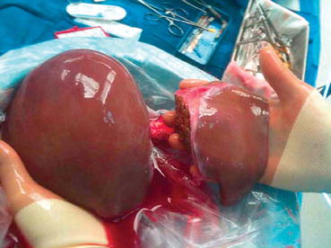
Split-liver cadaveric transplants are the least common type of transplant operation, accounting for only 1.0% of transplants performed in 2014 [].
Fig 11 Ex vivo split-liver cadaveric transplant the right tri-segment graft - photo 1
Fig. 1.1
Ex vivo split-liver cadaveric transplant the right tri-segment graft was transplanted into an adult recipient with HCC, while the left lateral segment graft went to an infant with fulminant hepatic failure. Adapted from Burk KS, Singh AK, Vagefi PA, Sahani D, Pretransplantation imaging workup of the liver donor and recipient. Radiology Clinics of North America 2016; 54(2):185197
Living Donor Liver Transplant
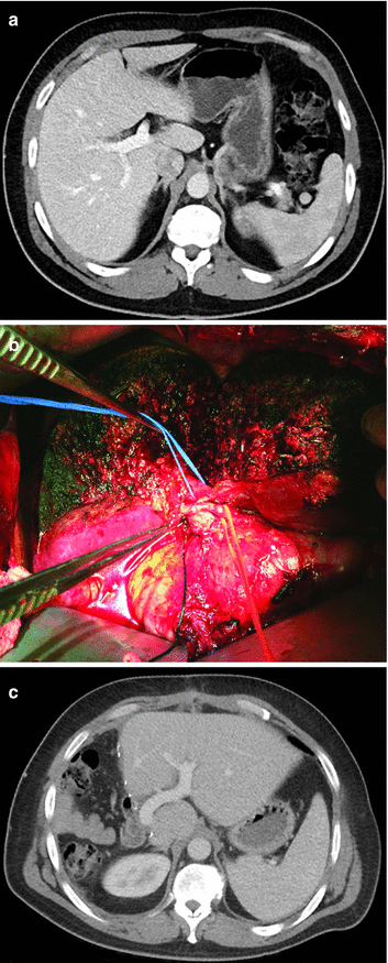
In a living donor liver transplant , a part of the donors liver is removed and transplanted into the recipient (Fig. ].
Fig 12 Right lobe living donor liver transplant a Preoperative donor - photo 2
Fig. 1.2
Right lobe living donor liver transplant. ( a ) Preoperative donor image. ( b ) Intraoperative photograph after liver parenchymal division but prior to vascular division. ( c ) Postoperative donor image showing growth in the remnant left hepatic lobe. Adapted from Burk KS, Singh AK, Vagefi PA, Sahani D, Pretransplantation imaging workup of the liver donor and recipient. Radiology Clinics of North America 2016; 54(2):185197
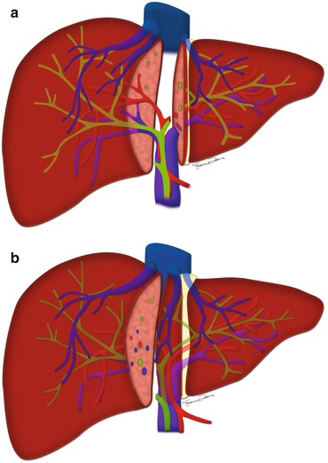
For a pediatric liver graft recipient, the left lateral segmentectomy technique is most popular. In this operation, the transection plane runs just to the right of the falciform ligament, and segment IV and the MHV are left in the donor (Fig. ].
Fig 13 a The left lateral segmentectomy LDLT plane runs to the left of - photo 3
Fig. 1.3
( a ) The left lateral segmentectomy LDLT plane runs to the left of the MHV. ( b ) The right lobe LDLT plane connects the gallbladder fossa and IVC and runs 1 cm to the right of the MHV. Adapted from Burk KS, Singh AK, Vagefi PA, Sahani D, Pretransplantation imaging workup of the liver donor and recipient. Radiology Clinics of North America 2016; 54(2):185197
The overall risk of postoperative complication for the partial liver donor is 40%, and the risk of postoperative death is 0.150.20%. Though this overall complication rate seems high, 95% of these complications are minor in severityClavien grade I or II. These require only conservative, medical management, or at most a percutaneous intervention [].
Imaging Techniques
CT and MRI are the modalities most commonly used to evaluate liver donors and recipients in the preoperative setting. Indeed, with the advent of biliary contrast agents and imaging post-processing techniques, older modalities including conventional angiography, ERCP, and intraoperative cholangiography have largely been replaced. Though MRI can be used as a sole imaging modality for preoperative evaluation, CT and MRI are more commonly used together as their strengths are complimentary to one another [].
CT/CTA
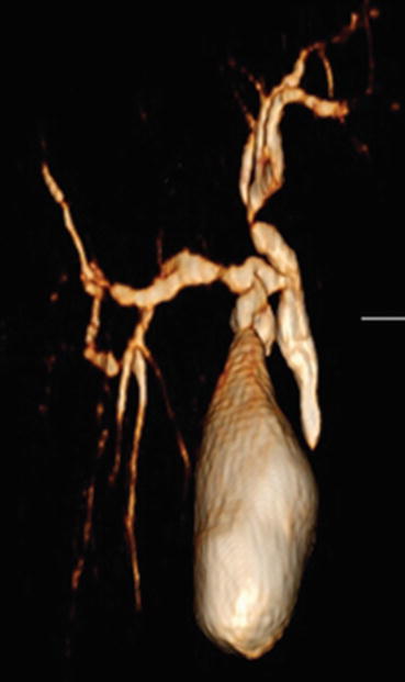
CT/CTA has superior spatial resolution compared to MRI and is therefore better at delineating small segmental hepatic arteries and accessory hepatic veins [). However, it is no longer performed as the biliary excreted iodinated contrast agent was removed from the US market a few years ago. Biliary evaluation is now performed via MRCP.
Fig 14 3D reformatted image of a CT cholangiogram showing the common bile - photo 4
Fig. 1.4
3D reformatted image of a CT cholangiogram showing the common bile duct and intrahepatic biliary ducts
Since liver donors are often young and healthy without other medical comorbidities, reduction in radiation dose for these exams is important. Methods to reduce radiation dose routinely used at our institution include limiting the field of view to the abdomen on arterial phase images, using iterative reconstruction and weight-based kVp techniques (80 kVp if <150 bs, 100 kVp if 150200 lbs, 120 kVp if >200 lbs) on portal/hepatic venous phase images, and using dual energy techniques (140 and 80 kVp) on only a limited number of thick slices (24 slices depending on liver size) for steatosis evaluation.
MRI/MRCP
MRI/MRCP with and without a hepatobiliary contrast agentGd-BOPTA (MultiHance) or Gd-EOB-DTPA (Eovist)is the second examination performed at our institution for evaluation of liver donors. Fat and iron deposition is assessed with Dixon sequences. The biliary system is evaluated with traditional T2-weighted non-contrast-enhanced 3D MRCP images in the coronal plane, T2-weighted SSFSE images in the coronal and axial planes, T1-weighted post-contrast biliary-phase images in the coronal and axial planes, and/or a 20-min delayed post-contrast 3D MRCP. Additionally, focal liver lesions are characterized with traditional T2, T1 pre-contrast, and T1 post-contrast images in the arterial, portal venous, and delayed phases []. For all the above, fast pulse sequences such as spoiled gradient echo and parallel imaging techniques are utilized to decrease image acquisition time and minimize motion artifacts.
Next page
Light

Font size:

Reset

Interval:

Bookmark:

Make

Similar books «Transplantation Imaging»

Look at similar books to Transplantation Imaging. We have selected literature similar in name and meaning in the hope of providing readers with more options to find new, interesting, not yet read works.


Reviews about «Transplantation Imaging»

Discussion, reviews of the book Transplantation Imaging and just readers' own opinions. Leave your comments, write what you think about the work, its meaning or the main characters. Specify what exactly you liked and what you didn't like, and why you think so.