Thomas H. Berquist - Imaging of Orthopaedic Fixation Devices and Prostheses
Here you can read online Thomas H. Berquist - Imaging of Orthopaedic Fixation Devices and Prostheses full text of the book (entire story) in english for free. Download pdf and epub, get meaning, cover and reviews about this ebook. year: 2012, publisher: Lippincott Williams & Wilkins, genre: Home and family. Description of the work, (preface) as well as reviews are available. Best literature library LitArk.com created for fans of good reading and offers a wide selection of genres:
Romance novel
Science fiction
Adventure
Detective
Science
History
Home and family
Prose
Art
Politics
Computer
Non-fiction
Religion
Business
Children
Humor
Choose a favorite category and find really read worthwhile books. Enjoy immersion in the world of imagination, feel the emotions of the characters or learn something new for yourself, make an fascinating discovery.
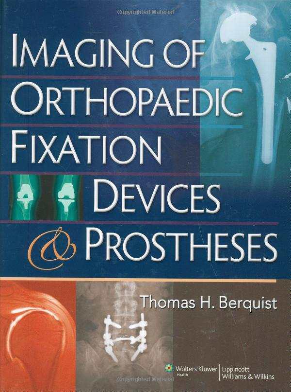
- Book:Imaging of Orthopaedic Fixation Devices and Prostheses
- Author:
- Publisher:Lippincott Williams & Wilkins
- Genre:
- Year:2012
- Rating:5 / 5
- Favourites:Add to favourites
- Your mark:
Imaging of Orthopaedic Fixation Devices and Prostheses: summary, description and annotation
We offer to read an annotation, description, summary or preface (depends on what the author of the book "Imaging of Orthopaedic Fixation Devices and Prostheses" wrote himself). If you haven't found the necessary information about the book — write in the comments, we will try to find it.
Note: This book is a condensed version of Berquists Imaging Atlas of Orthopaedic Appliances and Prostheses, which published in 1994.
Thomas H. Berquist: author's other books
Who wrote Imaging of Orthopaedic Fixation Devices and Prostheses? Find out the surname, the name of the author of the book and a list of all author's works by series.


Imaging of Orthopaedic Fixation Devices and Prostheses — read online for free the complete book (whole text) full work
Below is the text of the book, divided by pages. System saving the place of the last page read, allows you to conveniently read the book "Imaging of Orthopaedic Fixation Devices and Prostheses" online for free, without having to search again every time where you left off. Put a bookmark, and you can go to the page where you finished reading at any time.
Font size:
Interval:
Bookmark:
Acquisitions Editor: Lisa McAllister
Managing Editor: Ryan Shaw
Project Manager: Alicia Jackson
Manufacturing Coordinator: Kathleen Brown
Senior Marketing Manager: Angela Panetta
Designer: Stephen Druding
Cover Designer: Larry Didona
Production Services: Laserwords Private Limited, Chennai, India
2009 by LIPPINCOTT WILLIAMS & WILKINS, a WOLTERS KLUWER business
530 Walnut Street
Philadelphia, PA 19106 USA
LWW.com
All rights reserved. This book is protected by copyright. No part of this book may be reproduced in any form by any means, including photocopying, or utilized by any information storage and retrieval system without written permission from the copyright owner, except for brief quotations embodied in critical articles and reviews. Materials appearing in this book prepared by individuals as part of their official duties as U.S. government employees are not covered by the above-mentioned copyright.
Printed in China
Library of Congress Cataloging-in-Publication Data
Imaging of orthopaedic fixation devices and prostheses / editor, Thomas H. Berquist.
p. ; cm.
Includes bibliographical references and index.
ISBN-13: 978-0-7817-9252-3 (alk. paper)
ISBN-10: (invalid) 0-7817-9252-3 (alk. paper)
1. Orthopedic apparatusImaging. 2. Musculoskeletal systemDiseasesImaging. 3. Musculoskeletal system DiseasesSurgery. I. Berquist, Thomas H. (Thomas Henry), 1945-[DNLM: 1. Musculoskeletal Diseasesdiagnosis. 2. Diagnostic Imaging. 3. Musculoskeletal Diseasessurgery.
4. Orthopedic Fixation Devices. WE 141 I301 2009]
RD755.5.I38 2009
617'.9dc22
2008024236
Care has been taken to confirm the accuracy of the information presented and to describe generally accepted practices. However, the authors, editors, and publisher are not responsible for errors or omissions or for any consequences from application of the information in this book and make no warranty, expressed or implied, with respect to the currency, completeness, or accuracy of the contents of the publication. Application of this information in a particular situation remains the professional responsibility of the practitioner.
The authors, editors, and publisher have exerted every effort to ensure that drug selection and dosage set forth in this text are in accordance with current recommendations and practice at the time of publication. However, in view of ongoing research, changes in government regulations, and the constant flow of information relating to drug therapy and drug reactions, the reader is urged to check the package insert for each drug for any change in indications and dosage and for added warnings and precautions. This is particularly important when the recommended agent is a new or infrequently employed drug.
Some drugs and medical devices presented in this publication have Food and Drug Administration (FDA) clearance for limited use in restricted research settings. It is the responsibility of health care providers to ascertain the FDA status of each drug or device planned for use in their clinical practice.
To purchase additional copies of this book, call our customer service department at (800) 638-3030 or fax orders to (301) 223-2320. International customers should call (301) 223-2300.
Visit Lippincott Williams & Wilkins on the Internet: at LWW.com. Lippincott Williams & Wilkins customer service representatives are available from 8:30 am to 6 pm, EST.
10 9 8 7 6 5 4 3 2 1
I dedicate this text to my loving wife, Mary, for her continued support and understanding.

In 1995 we published an Atlas of Orthopaedic Appliances and Prostheses. This was a work dedicated to bridging the gap between orthopaedic surgeons and imagers. I have continued to dedicate efforts to improve the understanding of orthopaedic procedures and what the surgeon needs to know when ordering preoperative and postoperative imaging studies.
Orthopaedic instrumentation and prostheses continue to evolve, making it difficult for imagers to keep up with all possible implants that may appear on radiographs or other imaging modalities. With this in mind, it is essential for surgeons and radiologists to work closely and we, as imagers, need to become familiar with the instrumentation systems our surgeons prefer.
This edition is designed to be more concise than the prior atlas with no attempt to demonstrate every possible fixation device or prostheses. We review the important clinical and image features of orthopaedic devices including clinical concepts and patient selection, the normal appearance of orthopaedic devices and the image features, and most appropriate modalities for evaluating complications.
reviews clinical data, staging, and preoperative and postoperative imaging in patients with musculoskeletal neoplasms.
This text will be most useful to practicing radiologists and radiologists in training. Other physicians who deal with orthopaedic problems will also find the information provided in this text extremely useful.

Preparation of this text required the support of numerous individuals and colleagues. I first wish to thank my colleagues in musculoskeletal imaging at Mayo Jacksonville, Laura Bancroft, Mark Kransdorf, and Jeffrey Peterson for their support and assistance in providing the necessary images needed to fulfill the mission of this text. I also want to thank my colleagues in orthopaedic surgery, Mark Broderson, Stephen Trigg, Cedric Ortiguera, Peter Murry, Mary OConnor, Kurtis Blasser, and Joseph Whalen for their consultative support.
Daniel Huber and John Hagen were instrumental in providing images and art required to demonstrate anatomy, normal and abnormal image features for devices described in this text. The vendors of orthopaedic devices were also very helpful in providing photographs and artwork to assist with demonstration of devices and their indications to use along with the images in this text.
Finally, I wish to thank Ryan Shaw, Lisa McAlliser, and Kerry Barrett from Lippincott Williams & Wilkins for their assistance and support with this project.


 ppropriate use of imaging techniques is essential for diagnosis, treatment planning, and follow-up of orthopaedic procedures. Basic techniques will be discussed in this chapter to avoid redundancy in anatomic chapters.
 Routine Radiographs
Routine radiographs remain the primary screening examination for musculoskeletal disorders. Appropriate evaluation of radio-graphs may provide the diagnosis or allow selection of the next imaging procedure to completely evaluate the clinical problem. Specifically, radiographs are essential for proper interpretation of magnetic resonance (MR) images.
Currently, screen-film radiography is being replaced with computed radiography (CR) at many institutions. Regardless of the system used, it is essential to ensure proper patient positioning and accurate chronologic labeling of images. Multiple views (two to four) are required to evaluate osseous and articular anatomy. Specific views will be discussed in subsequent anatomic chapters. In certain cases, fluoroscopically positioned images are useful to optimize positioning and reduce bony overlap. This approach is useful in the foot and wrist. The technique is also appropriate to evaluate interfaces of arthroplasty components, fixation devices, and evaluate pin tracts when infection is suspected clinically. Fluoroscopic positioning is also useful when performing stress tests to assure that the joint is properly positioned. Stress studies are most often performed on the ankle, elbow, knee, and wrist (see ).
Font size:
Interval:
Bookmark:
Similar books «Imaging of Orthopaedic Fixation Devices and Prostheses»
Look at similar books to Imaging of Orthopaedic Fixation Devices and Prostheses. We have selected literature similar in name and meaning in the hope of providing readers with more options to find new, interesting, not yet read works.
Discussion, reviews of the book Imaging of Orthopaedic Fixation Devices and Prostheses and just readers' own opinions. Leave your comments, write what you think about the work, its meaning or the main characters. Specify what exactly you liked and what you didn't like, and why you think so.