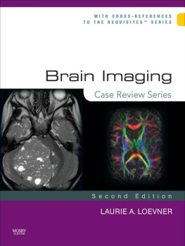
Case-Based Brain Imaging
Second Edition
A. John Tsiouris, MD
Associate Professor of Clinical Radiology
Weill Cornell Medical College
NewYork-Presbyterian Hospital
New York, New York
Pina C. Sanelli, MD, MPH
Associate Professor of Radiology and Public Health
Weill Cornell Medical College
NewYork-Presbyterian Hospital
New York, New York
Joseph P. Comunale, MD
Associate Professor of Clinical Radiology
Weill Cornell Medical College
NewYork-Presbyterian Hospital
New York, New York
Thieme
New York Stuttgart
Thieme Medical Publishers, Inc.
333 Seventh Ave.
New York, New York 10001
Executive Editor: Timothy Hiscock
Managing Editor: J. Owen Zurhellen IV
Editorial Assistant: Elizabeth Berg
Senior Vice President, Editorial and Electronic Product Development: Cornelia Schulze
Production Editor: Heidi Grauel, Maryland Composition
International Production Director: Andreas Schabert
Vice President, Finance and Accounts: Sarah Vanderbilt
President: Brian D. Scanlan
Compositor: Maryland Composition
Printer: Everbest Printing Co.
Library of Congress Cataloging-in-Publication Data
Case-based brain imaging/edited by A. John Tsiouris, Pina C. Sanelli, Joseph P. Comunale.2nd ed.
p.; cm.
Rev. ed. of: Teaching atlas of brain imaging/Nancy J. Fischbein, William P. Dillon, A. James Barkovich.
2000.
Includes bibliographical references and index.
ISBN 978-1-60406-953-2
I. Tsiouris, A. John. II. Sanelli, Pina C. III. Comunale, Joseph P. IV. Fischbein, Nancy J. Teaching atlas of brain imaging.
[DNLM: 1. Brain DiseasesdiagnosisAtlases. 2. Brain DiseasesdiagnosisCase Reports.
3. Diagnostic ImagingAtlases. 4. Diagnostic ImagingCase Reports. WL 17]
616.80475dc23
2012039003
Copyright 2013 by Thieme Medical Publishers, Inc. This book, including all parts thereof, is legally protected by copyright. Any use, exploitation, or commercialization outside the narrow limits set by copyright legislation without the publishers consent is illegal and liable to prosecution. This applies in particular to photostat reproduction, copying, mimeographing or duplication of any kind, translating, preparation of microfilms, and electronic data processing and storage.
Important note: Medical knowledge is ever-changing. As new research and clinical experience broaden our knowledge, changes in treatment and drug therapy may be required. The authors and editors of the material herein have consulted sources believed to be reliable in their efforts to provide information that is complete and in accord with the standards accepted at the time of publication. However, in view of the possibility of human error by the authors, editors, or publisher of the work herein or changes in medical knowledge, neither the authors, editors, nor publisher, nor any other party who has been involved in the preparation of this work, warrants that the information contained herein is in every respect accurate or complete, and they are not responsible for any errors or omissions or for the results obtained from use of such information. Readers are encouraged to confirm the information contained herein with other sources. For example, readers are advised to check the product information sheet included in the package of each drug they plan to administer to be certain that the information contained in this publication is accurate and that changes have not been made in the recommended dose or in the contraindications for administration. This recommendation is of particular importance in connection with new or infrequently used drugs.
Some of the product names, patents, and registered designs referred to in this book are in fact registered trademarks or proprietary names even though specific reference to this fact is not always made in the text. Therefore, the appearance of a name without designation as proprietary is not to be construed as a representation by the publisher that it is in the public domain.
Printed in China
5 4 3 2 1
ISBN 978-1-60406-953-2
eISBN 978-1-60406-954-9
To our patients, who are an infinite source of challenging cases that motivate us to continuously improve our knowledge and skills.
I dedicate this book to my father, Dr. John A. Tsiouris, for the many sacrifices he made throughout his life so my brother and I could succeed.
Apostolos John Tsiouris, MD
I dedicate this book to my loving and supportive husband, George, and to our three children, Isabella, Sophia, and Nicholas, who are truly our pride and joy.
Pina C. Sanelli, MD, MPH
I dedicate this book to my parents, for their unconditional support and encouragement, and to my colleagues, residents, and fellows, who continue to motivate me to be the best I can be.
Joseph P. Comunale, MD
To access additional material or resources available with this e-book, please visit http://www.thieme.com/bonu scontent. After completing a short form to verify your e-book purchase, you will be provided with the instructions and access codes necessary to retrieve any bonus content.
Contents
by Robert D. Zimmerman, MD, FACR
Foreword
The second edition of the popular Teaching Atlas of Brain Imaging by Drs. Fischbein, Dillon and Barkovich has finally been produced after a hiatus of 12 years, now renamed Case-Based Brain Imaging. The wait was clearly worth it!
The editors of this second edition, Drs. Tsiouris, Comunale, and Sanelli, are all my colleagues at NewYork-Presbyterian, Weill Cornell Medical College. They are outstanding clinicians and teachers who have used their combined experience and expertise to carefully choose 152 first-rate CT and MRI cases that illustrate the key imaging features of the full spectrum of brain disease in an easy-to-access format. The result is a book that is both comprehensive and concise. Each chapter starts with an unknown case. In many of the chapters, additional companion images and cases are provided to enhance the readers knowledge of the topic and demonstrate variations of the profiled disease. The key imaging, pathologic, and pathophysiologic findings for each disease are clearly outlined for each case. I especially appreciate the Pearls and Pitfalls sections at the end of each case that summarize wise tips for the reader.
Since the first edition, there have been major advances in the CT and MR imaging techniques utilized in neuroradiology. Numerous cases in this text include imaging techniques such as CT angiography, MR angiography, CT and MR perfusion, and MR spectroscopy that are now commonly used in practice for the diagnosis and surveillance of CNS disease. The all-new images are spectacular, having been obtained on state-of-the-art CT and high field MR scanners. The discussions are clear and concise, and the references have all been updated and the cases presented in a clean and uncluttered layout.
This book is meant to provide trainees and practicing radiologists, neurologists, and neurosurgeons with an opportunity to learn quickly about entities they encounter in their daily clinical practice, and it succeeds in this mission admirably. If you see it in practice, it is included in this book. It also includes numerous rare zebra cases that can cause diagnostic dilemmas. Lastly, this excellent text provides the reader with the opportunity to test their skills in the interpretation of unknown cases. For me this is the guilty pleasure of this book. Lets face it, radiologists love visual puzzles. The enduring popularity of case-of-the-day presentations, unknown case sessions, and film panels at our national meetings speaks to this love. This book offers each of us the opportunity to test our knowledge on representative cases and, in the process, gain significant information about a variety of entities.
Next page
