Springer-Verlag Italia 2016
Fabio Triulzi , Cristina Baldoli , Cecilia Parazzini and Andrea Righini Perinatal Neuroradiology 10.1007/978-88-470-5325-0_1
1. Normal Development
Fabio Triulzi 1 , Elisa Scola 1 and Sabrina Avignone 1
(1)
Department of Neuroradiology, Fondazione IRCCS C Granda, Ospedale Maggiore Policlinico, Milan, Italy
Fetal MR imaging covers a relatively long period of the fetal brain development: from approximately 1819 gestational weeks (GW) until birth. Therefore, at present, it is possible to study more than ahalf of the entire gestational period by this technique.
Different authors and most of all,Catherine Garel,have reviewed systematically the normal development of the fetal brain as it appears on the MR images and reported normal biometrics curves as measured by MRI;gyration and myelination process has been extensively reported as well[].
Here we present the fetal brain normal anatomy by means of MRI, with particular focus on the crucial period between 19 and 22 GW, taking into accountthe high-resolutionimages of MR autopsy (Fig. ) as reference guideline to interpret the low-resolution fetal MR images.
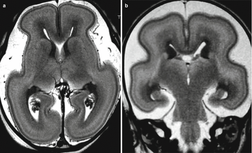
Fig. 1.1
Fetal MR autopsy. Normal brain at 22 gestational weeks (GW) ( a ) and 21 GW ( b ) on T2-weighted images. Voxel size 100 nl.
1.1 Fetal MR Autopsy Technique
In order to prevent postmortem tissue autolysis, fetal MR autopsy studies were carried out within 24 h from death, without any fixation, in an intact fetus conserved in a refrigerator at 45C prior to MR examination. The fetuses withspontaneous death in utero were not considered due to their long permanenceat body temperature that accelerates the autolysis.
To obtain comparable images with the in vivo study, it is important to preserve the natural tissue contrast on T1 and T2-weighted images. The standard fixation with formalin causes a marked tissue dehydration and a modification of T1 and T2 contrast [].
To obtain a reasonable compromise between acquisition time in a clinical setting and spatial resolution, the total acquisition time of a fetal MR autopsy is approximately 80 min by using a3.0 T magnet. The T2-weighted images shown in this chapter are obtained applying a turbo spin-echo sequence (TR 6500 ms; TE 120 ms; FA 90; NEX 4,acquisition time 20 min.); the voxel size was 0.30.31.2 mm, equal to a true spatial resolution of 0.10 mm3 (100 nl).
1.2 Fetal Anatomy on MR Autopsy
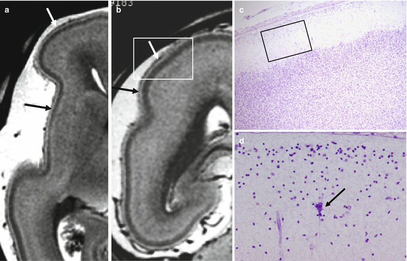
On the T2-weighted images of the fetal MR autopsy with spatial resolution of 100 nl, not only thethree layers usually recognizable on in vivo fetal MR are visible, but also layer number 1, the marginal layer (Fig. ], but at present, no correlation between these images and corresponding pathological specimens is available.
Fig. 1.2
Marginal zone on fetal MR autopsy (22GW). Layer I or marginal zone is detectable on fetal MR autopsy with a voxel size of 100 nl ( arrows a , b ). Correspondent histologic section stained with hematoxylineosin ( c ) and closeup view on marginal zone shows a large CajalRetzius cell ( black arrow ) ( d ) (courtesy C. Frassoni, Neurological Institute C. Besta, Milan)
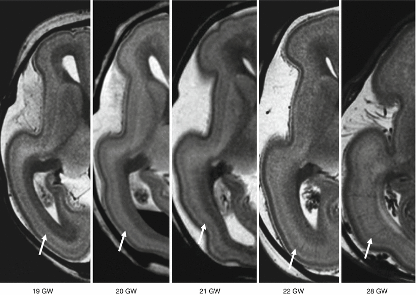
Fig. 1.3
A diffuse T2 hypointense layer on the more external part of subplate is recognizable on fetal MR autopsy between 19 and 28 GW ( arrows ). According to Kostovic et al., it could represent thalamocortical axons in the superficial layer of subplate that are waiting to enter the cortical plate
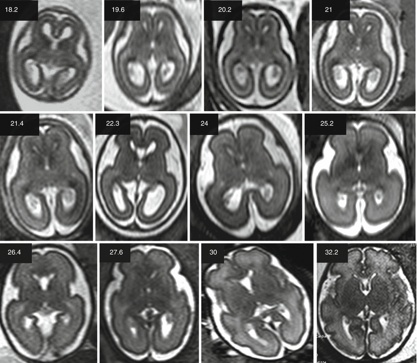
If we compare the single-shot T2-weighted images of the in vivo fetal MR (Fig. ).
Fig. 1.4
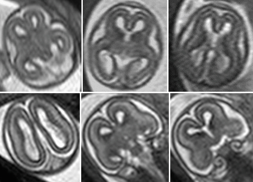
Normal fetal brain development according to fetal MR. Twelve selected cases from 18.2 to 32.2 GW, axial T2-weighted images passing approximately through the thalamus. For the acquisition technique, see the text
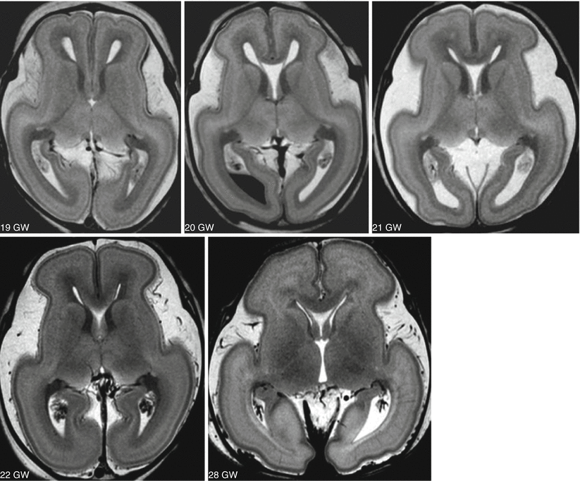
Fig. 1.5
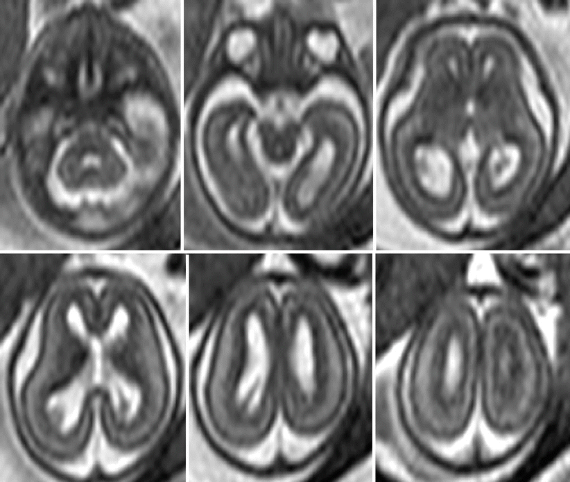
Fetal MR autopsy. Normal cases at 19, 20, 21, 22, and 28 GW, representative axial T2-weighted images passing through the thalamus. Voxel size 100 nl. For the acquisition technique, see the text; all the cases are studied within 24 hours from termination of pregnancy ( TOP )
Fig. 1.6
Fetal MR, normal brain at 18.2 GW. Fetal MR is usually not performed before 19 GW. The fetal brain size is too small and the fetal movement is more pronounced. In this case, the parenchymal thickness is still relatively thin in comparison to the ventricular size ( top row ). T2 signal contrast differences between cortical plate, subplate, and intermediate zone are however already appreciable ( bottom row )
Fig. 1.7
Fetal MR, normal brain at 19.6 GW, axial sections. At this age, the main brain mantle layers, including germinal matrix, are usually well documented. Brain opercularization is going to become evident even though the surface of the brain is still substantially smooth
Fig. 1.8
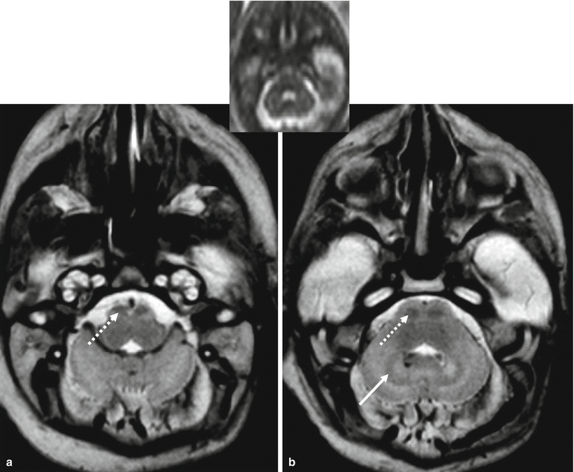
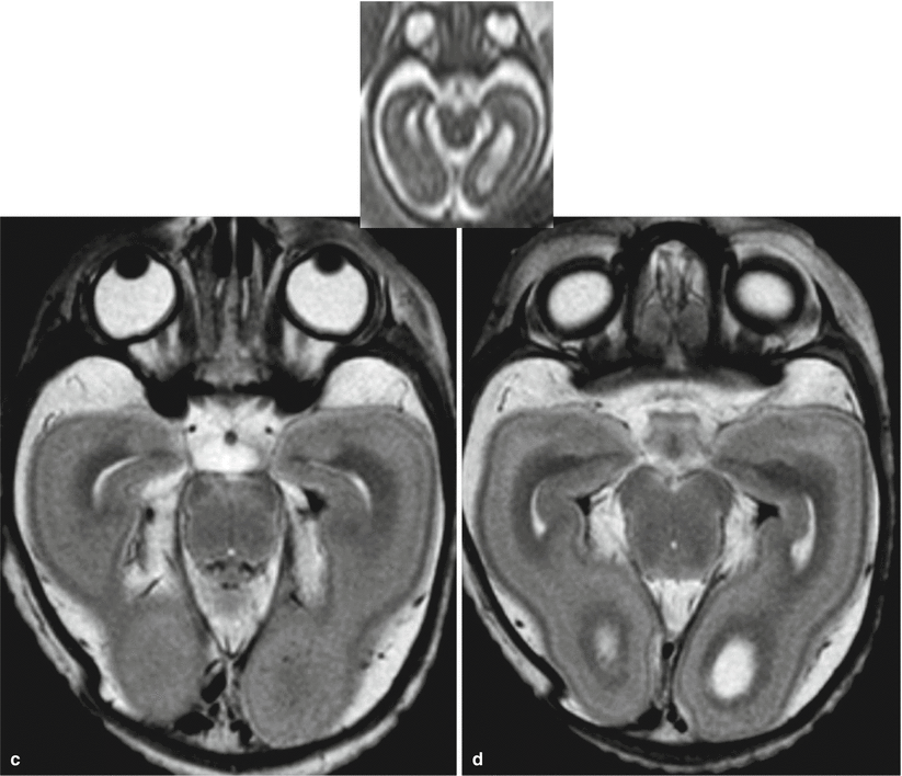
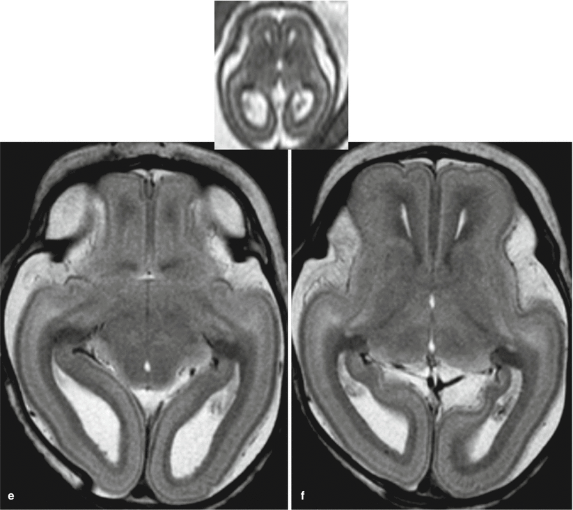
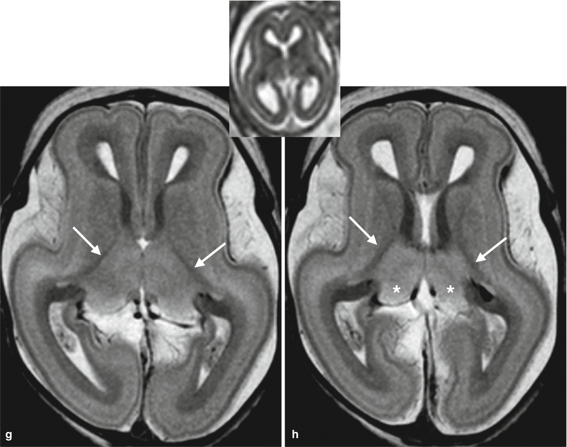
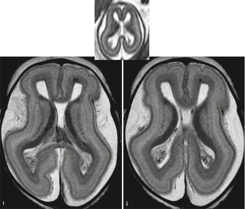
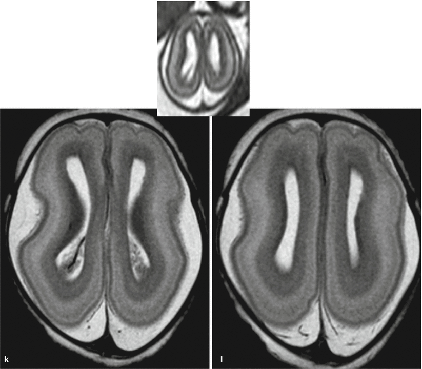
Fetal MR autopsy, normal brain at 19 GW, axial sections with correspondent fetal MR in vivo axial sections. Even without an appreciable myelination, corticospinal tracts are recognizable as relatively T2 hypointense bilateral symmetric areas in the most antero-mesial part of the medulla oblongata ( dotted arrow a ) and as a small oval areas in the anterior pons ( dotted arrow b ). Similarly, the posterior limb of the internal capsule is already present, and even without myelin, the fibers appear as relatively hypointense structure with respect to the basal ganglia and thalamus ( arrows g h ). The posterior part of the thalami, mainly represented by the pulvinar, is relatively hyperintense on T2-weighted images ( asterisk g h ). Cerebellar hemispheres are still small, but dentate nuclei are already visible ( b arrow ). Eye globes show the typical immature aspect with an irregular oval morphology ( c ). Germinal matrix is clearly visible as thethick most hypointense layer along the median portion of the lateral ventricles, adiacent to the forming basal ganglia ( g k ). Temporal lobes opercularization is going to be visible ( d h ). The three major layers, cortical plate, subplate, and intermediate zone, are easily visible; marginal zone or layer I is also visible as well as the hypointense layer in the outer part of the subplate, likely to becompatible with thalamocortical axons. The irregular indentations of cortical plate at the level of frontal lobes are postmortem artifacts ( f l )