Apostolos H. Karantanas (ed.) Medical Radiology Sports Injuries in Children and Adolescents 10.1007/174_2010_6 Springer-Verlag Berlin Heidelberg 2010
Common Injuries in Gymnasts
Maaike P. Terra 1, Mario Maas 1 , Charlotte M. Nusman 1, Ana Navas-Canete 1 and Milko C. de Jonge 1
(1)
Department of Radiology, Academic Medical Center, Meibergdreef 9, 1105 AZ Amsterdam, The Netherlands
2.1
2.1.1
2.1.2
2.2
2.2.1
2.2.2
2.2.3
2.3
2.3.1
2.3.2
2.3.3
3.1
3.1.1
3.1.2
3.1.3
3.1.4
3.2
3.2.1
3.2.2
3.3
3.3.1
3.3.2
3.3.3
4.1
4.2
4.3
Abstraxt
Both acute injuries as well as chronic overuse injuries should be considered when evaluating examinations of elite gymnastic athletes. Injuries of the growing skeleton are typically seen in gymnasts. Imaging in gymnasts comprises mainly conventional radiographs, MR imaging and CT. There is an important additional role for Multi detector Helical CT when MR imaging is suggestive of disease: CT will better delineate extend of lesion especially in the osseous structures. Correlation between imaging findings and clinical situation is mandatory. Therefore close interaction between radiologist and treating physician is essential.
Key Points
Both acute injuries as well as chronic overuse injuries should be considered when evaluating examinations of elite gymnastic athletes.
Injuries of the growing skeleton are typically seen in gymnasts.
Imaging in gymnasts comprises mainly conventional radiographs, MR imaging and CT.
There is an important additional role for Multi detector Helical CT when MR imaging is suggestive of disease: CT will better delineate extend of lesion especially in the osseous structures.
Correlation between imaging findings and clinical situation is mandatory. Therefore close interaction between radiologist and treating physician is essential.
Introduction
Participation in gymnastics has undergone rapid growth in past decades, especially in the female population (Backx et al. ).
Gymnastics-related injuries can roughly be divided in two main categories, acute injuries and chronic overuse problems. Acute injuries, usually resulting from a fall or faulty landing, comprise primarily of fractures, dislocations, sprains and strains. Chronic overuse problems result from chronic repetitive injury over an extended period of time leading to e.g., stress fractures and osteochondral injuries. Caine and Nassar revealed in a recent review of the literature that the majority of injuries in gymnastics were of sudden onset in nature (range = 5283.4%) (Caine and Nassar ). They also showed that the pattern of injuries varies according to the competition level, with more chronic injuries found in elite compared to nonelite gymnasts. Injuries also vary by location. The ankle injuries are mainly due to an acute trauma whereas wrist and low back injuries are generally due to chronic overuse.
In this chapter an overview is given of typical gymnastics-related injuries of the upper extremities, lower extremities and the axial skeleton in elite gymnasts. To prevent overlap with other chapters we have focused per category only on the most common injuries and their radiological features as seen in our institution.
Upper Extremity Injuries
During gymnastic activities the upper extremity is generally used to support body weight and is subjected to many different types of stress, including repetitive motion, high impact loading and axial compression. A review of upper extremity injuries in gymnasts showed that the highest incidence occurs in the wrist in females and the shoulder in male gymnasts (Caine and Nassar ).
2.1 Wrist
2.1.1 Growth Plate Injury
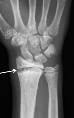
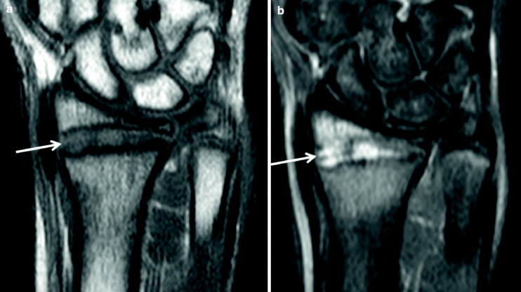
In the immature skeleton, the growth plates of the wrist are at risk for injury. Because most of the load applied to the wrist in dorsal flexion is applied to the distal radius, most of the overuse injuries are found in the distal radial physis with prevalence rates ranging from 10 to 85% (Caine and Nassar ).
Fig. 1
The PA radiograph of the right wrist in a 14-year-old female gymnast, demonstrates widening of the growth plate of the distal radius with lucent areas mainly at the metaphyseal aspect of the growth plate ( white arrow )
Fig. 2
Coronal T1-w ( a ) and STIR ( b ) MR images from the same patient as Fig.. Widening of the growth plate with an irregular surface of the metaphyseal aspect of the growth plate and vertical fractures is best seen at the T1-w image ( white arrow ), while the hyperintense signal of the widened physeal plate with the associated epiphyseal and metaphyseal bone marrow edema is best visualized at the STIR image ( white arrow )
2.1.2 Fracture
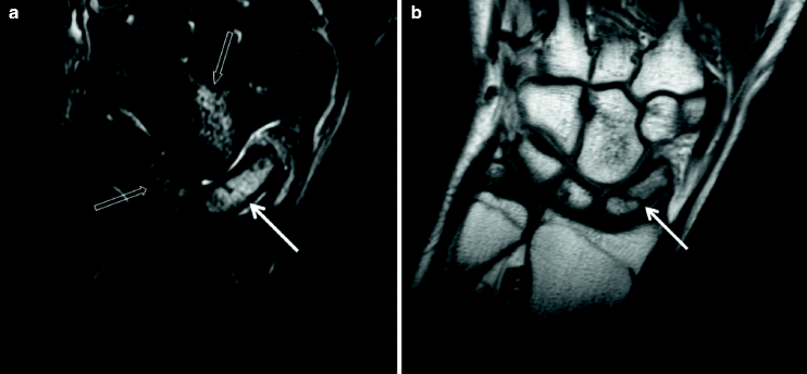
The most frequent, and also the most problematical fracture, in the wrist of gymnasts is that of the scaphoid. Scaphoid fractures in general account for approximately 60% of all carpal fractures. They occur primarily during a fall on the outstretched hand. However, in gymnastics, extreme dorsiflexion drives the scaphoid volarly and repetitive forces applied to it may lead to a stress fracture. Both acute and stress fractures occur most commonly at the waist (7080%), the weakest point in the scaphoid, but the fracture line can also be located in the tuberosity, the distal pole (510%) or the proximal pole (1520%).
Posteroanterior and lateral radiographs of the wrist together with scaphoid-specific views usually suffice to detect the fracture line in acute injuries. However, initial radiographs may be negative and in these cases MR imaging or multi-detector CT can be considered (Karantanas et al. ). In gymnasts with a scaphoid stress fracture, radiographs may initially show sclerosis at the waist and later a discrete fracture line.
Fig. 3
The coronal STIR ( a ) and T1-w ( b ) MR images in a 16-year-old female patient who sustained a fall during training, demonstrate bone marrow edema of the scaphoid ( a , white arrow ) with a proximal pole fracture ( b , white arrow ). Also bone marrow edema (bone bruise) in the absence of a fracture is shown in the capitate and lunate ( a , open white arrows )
2.2 Elbow
2.2.1 Osteochondritis Dissecans
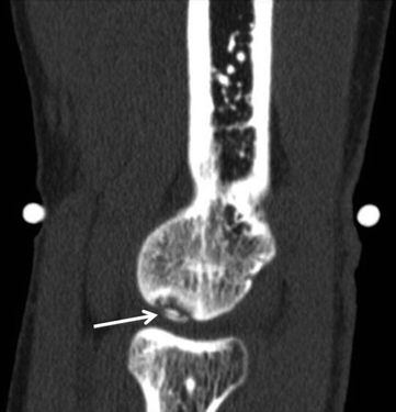
Osteochonditis dissecans (OD) is a form of osteochondrosis of the articular epiphysis. In the elbow of the gymnasts this condition is frequently seen at the humeral capitellum and trochlea, mainly in females (Bradley and Petrie ).
Fig. 4
Sagittal CT MPR image showing an osteochondral lesion with a loose bone fragment of the capitellum ( arrow ) in a 15-year-old female gymnast
