• Complain

Pu Dai Dong-yi Han Vincent C Cousins - Stereo Operative Atlas of Micro Ear Surgery

Here you can read online Pu Dai Dong-yi Han Vincent C Cousins - Stereo Operative Atlas of Micro Ear Surgery full text of the book (entire story) in english for free. Download pdf and epub, get meaning, cover and reviews about this ebook. City: Singapore, year: 2018, publisher: Springer Singapore;SPRINGER Verlag, SINGAPOR, genre: Home and family. Description of the work, (preface) as well as reviews are available. Best literature library LitArk.com created for fans of good reading and offers a wide selection of genres:

Romance novel Science fiction Adventure Detective Science History Home and family Prose Art Politics Computer Non-fiction Religion Business Children Humor

Choose a favorite category and find really read worthwhile books. Enjoy immersion in the world of imagination, feel the emotions of the characters or learn something new for yourself, make an fascinating discovery.

Pu Dai Dong-yi Han Vincent C Cousins Stereo Operative Atlas of Micro Ear Surgery

Stereo Operative Atlas of Micro Ear Surgery: summary, description and annotation

We offer to read an annotation, description, summary or preface (depends on what the author of the book "Stereo Operative Atlas of Micro Ear Surgery" wrote himself). If you haven't found the necessary information about the book — write in the comments, we will try to find it.

Pu Dai Dong-yi Han Vincent C Cousins: author's other books


Who wrote Stereo Operative Atlas of Micro Ear Surgery? Find out the surname, the name of the author of the book and a list of all author's works by series.

Stereo Operative Atlas of Micro Ear Surgery — read online for free the complete book (whole text) full work

Below is the text of the book, divided by pages. System saving the place of the last page read, allows you to conveniently read the book "Stereo Operative Atlas of Micro Ear Surgery" online for free, without having to search again every time where you left off. Put a bookmark, and you can go to the page where you finished reading at any time.

Light

Font size:

Reset

Interval:

Bookmark:

Make
Springer Science+Business Media Singapore and People's Medical Publishing House 2017
Pu Dai , Dong-yi Han , Vincent C Cousins and Yue-shuai Song (eds.) Stereo Operative Atlas of Micro Ear Surgery 10.1007/978-981-10-2089-6_1
1. General Introduction
Yue-shuai Song 1
(1)
Department of Otolarygngology Head and Neck Surgery, Beijing Friendship Hospital, Capital Medical University, Beijing, 100050, China
(2)
Department of Otolaryngology, The 175th Hospital of Chinese PLA, Zhangzhou, 363000, China
(3)
Department of Otolarygngology Head and Neck Surgery, Chinese PLA General Hospital, Beijing, 100853, China
Yue-shuai Song (Corresponding author)
Email:
Pu Dai (Corresponding author)
Email:
A Brief History of the Development of Stereoscopy in Ear Micro Surgery
Yue-shuai Song 4 and Pu Dai 5
(4)
Department of Otolarygngology Head and Neck Surgery, Beijing Friendship Hospital, Capital Medical University, Beijing, 100050, China
(5)
Department of Otolarygngology Head and Neck Surgery, Chinese PLA General Hospital, Beijing, 100853, China
Our knowledge of the anatomy of the temporal bone can be dated back to Hippocrates who understood the tympanic membrane to be part of the acoustic apparatus, and Aristotle who recognized the cochlea as part of the hearing organ. Later in 1860 Toynbee studied 2000 temporal bones and published his findings in the classic work Disease of the Ear. So techniques especially modern techniques of dissection have greatly improved our abilities to study the temporal bone, and among them, Computerized Tomography (CT Scanning) has added to this.
The basic threads of three dimensional (3D) spatial shape research of otology came from Newton. In 1879 he suggested reconstructing the temporal bone using contiguous sheets, but this was not available until 1968, when an English engineer Hounsfield invented the first CT machine. With this technique Harada (1988) made some particular observations of the temporal bone, including the external auditory canal, tympanic membrane, carotid artery, vestibule, labyrinth and internal auditory canal. In 1989, Lutz reconstructed the facial nerve and carotid artery and made a 3-D measurement of anatomical structures. He also discussed the anatomical basis of various otologic diseases. In China, the work of Professor Pu Dai (1991) on temporal bone reconstruction based on extensive anatomic data hasnt only expanded the study but also enriched the knowledge of stereo reconstruction in China.
Fig 11 Reconstructed 3D cochlea and reflecting mirror stereoscope The - photo 1
Fig. 1.1
Reconstructed 3D cochlea and reflecting mirror stereoscope
The wheatstone stereoscope (1838): to view images using the stereoscope (Reprint from Volume 68 of the series Intelligent Systems, Control and Automation: Science and Engineering pp 97130, Springer, 2013)
Computer aided temporal bone reconstruction has become a landmark in Medical studies, while it still has some defects, the images which are created by this technique are only an abstraction of the real anatomic structures, far different to real surgical images. Considering this, Professor Dai has further developed a way to capture and save stereo images. He has combined binocular vision theory, the stereoscope and the operating microscope in an ingenious way. This has provided a much more realistic representation of micro ear surgery.
Stereo Operative Photography of Micro Ear Surgery
Song Gao 6 and Pu Dai 7
(6)
Department of Otolaryngology, The 175th Hospital of Chinese PLA, Zhangzhou, 363000, China
(7)
Department of Otolarygngology Head and Neck Surgery, Chinese PLA General Hospital, Beijing, 100853, China
What Is Stereo Images of Micro Ear Surgery and How to Capture Them?
Micro Ear Surgery is routinely performed with the help of the operating microscope which gives true stereo vision through the two eye lens (just like we watch things through our two naked eyes). Traditionally this level of stereo-vision has not been available outside the Operating Room and we have had to bear standard 2D pictures, losing a lot of important spatial information, but now we have used a side-by-side stereoscopic system to address this deficiency. We use two separate Digital Single Lens Reflex (SLR) Cameras attached to an operating microscope to simultaneously capture left and right pictures of the same object as shown in the picture below.
Equipment and Specifications
The following equipment is required to record the ideal paired photographs:
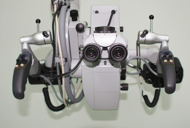
  1. Operating microscope with two side ports for attachment of two digital SLR cameras. Each side of the system, including eye-lens and camera, must have the same optical pathway;
  2. The two cameras should be controlled by one manual key or pedal so that the left and right photographs are taken simultaneously by two cameras separately;
  3. Adequate light: Sufficient light is always very important, especially when you want to capture images of very tiny structures in a very deep situation;
  4. Two Hi-Q Digital Single Lens Reflex Cameras, and make sure they share the same settings.
In our department, we use two Canon EOS 5D digital cameras, one Carl Zeiss Pentero microscope with a foot control pedal connecting to it, which is used for image capture. Correct focus is controlled via a video monitor that has the same optical pathway to the right eye-lens.
Fig 12 The microscope with stereoscopic photo system Stereo Image - photo 2
Fig. 1.2
The microscope with stereoscopic photo system
Stereo Image Production
  • Step 1: Open a pair of photos, captured during surgery in Adobe Photoshop software;
  • Step 2: Overlap the paired left and right side photos, make sure the structure in the focus of these two pictures can be completely matched.
  • Step 3: Crop photos to a standard measurement, and keep the observation target in the middle of the field;
  • Step 4: Position the edited photos side by side in their original side position;
  • Step 5: Save the side-by-side stereo-images in Hi-Q JPEG format;
The following steps are necessary to ensure effective stereovision:
  1. Make sure the structure in the focus of these two pictures can be completely overlapped;
  2. Side cannot be exchanged.
How to View the Stereo Images
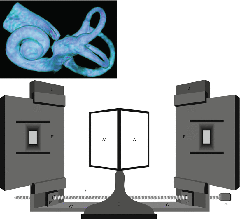
  • Step 1: Unfold the stereoscope;
  • Step 2: Place the two feet of stereoscope in the both lateral margins of side-by-side stereo-image;
  • Step 3: Adjust lens distance of stereoscope;
  • Step 4: Enjoy the realistic stereovision!
Suggested Reading
A Brief Development History of Stereoscopic in Micro Ear Surgery
Beckmann EC. CT scanning the early days. Br J Radiol. 2006;79(937):58. CrossRef PubMed
Dai P, Jiang SC, Gu R. Computer assisted three-dimensional (3D) reconstruction and mechanical model establishment of Ossicular chain. Chin J Otorhinolaryngol. 1991;26:2724.
Next page
Light

Font size:

Reset

Interval:

Bookmark:

Make

Similar books «Stereo Operative Atlas of Micro Ear Surgery»

Look at similar books to Stereo Operative Atlas of Micro Ear Surgery. We have selected literature similar in name and meaning in the hope of providing readers with more options to find new, interesting, not yet read works.


Reviews about «Stereo Operative Atlas of Micro Ear Surgery»

Discussion, reviews of the book Stereo Operative Atlas of Micro Ear Surgery and just readers' own opinions. Leave your comments, write what you think about the work, its meaning or the main characters. Specify what exactly you liked and what you didn't like, and why you think so.