Renato Hoffmann Nunes Ana Lorena Abello - Critical Findings in Neuroradiology
Here you can read online Renato Hoffmann Nunes Ana Lorena Abello - Critical Findings in Neuroradiology full text of the book (entire story) in english for free. Download pdf and epub, get meaning, cover and reviews about this ebook. City: Cham, year: 2018, publisher: Springer International Publishing, genre: Home and family. Description of the work, (preface) as well as reviews are available. Best literature library LitArk.com created for fans of good reading and offers a wide selection of genres:
Romance novel
Science fiction
Adventure
Detective
Science
History
Home and family
Prose
Art
Politics
Computer
Non-fiction
Religion
Business
Children
Humor
Choose a favorite category and find really read worthwhile books. Enjoy immersion in the world of imagination, feel the emotions of the characters or learn something new for yourself, make an fascinating discovery.

- Book:Critical Findings in Neuroradiology
- Author:
- Publisher:Springer International Publishing
- Genre:
- Year:2018
- City:Cham
- Rating:3 / 5
- Favourites:Add to favourites
- Your mark:
Critical Findings in Neuroradiology: summary, description and annotation
We offer to read an annotation, description, summary or preface (depends on what the author of the book "Critical Findings in Neuroradiology" wrote himself). If you haven't found the necessary information about the book — write in the comments, we will try to find it.
Renato Hoffmann Nunes Ana Lorena Abello: author's other books
Who wrote Critical Findings in Neuroradiology? Find out the surname, the name of the author of the book and a list of all author's works by series.


Critical Findings in Neuroradiology — read online for free the complete book (whole text) full work
Below is the text of the book, divided by pages. System saving the place of the last page read, allows you to conveniently read the book "Critical Findings in Neuroradiology" online for free, without having to search again every time where you left off. Put a bookmark, and you can go to the page where you finished reading at any time.
Font size:
Interval:
Bookmark:
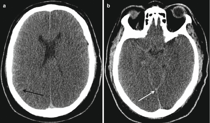
Brain
- Focal : Generates a pressure gradient in adjacent regions and may result in tissue shifts and herniations. Examples of focal edema can be found around tumors, hematomas, and infarctions [].
- Global : Diffusely affects the whole brain and, when critical, may cause intracranial hypertension and compromised perfusion and lead to generalized ischemia. Cardiopulmonary arrest, severe traumatic injury, multisystem organ failure, hypertensive crisis, infection or inflammation, hypoxicischemic injury, and toxic and metabolic conditions are common causes of global cerebral edema [].
Cytotoxic |
Arterial infarction |
Small vessel disease |
Vasogenic |
Neoplasm |
Hemorrhage |
Venous thrombosis |
Arteriovenous shunts |
Interstitial |
Hydrocephalus |
Combined |
Trauma |
Hypoxicischemic encephalopathy |
Osmotic |
Hydrostatic |
Infection or inflammation |
- Cytotoxic edema : Cytotoxic edema is defined as a cellular process, where extracellular Na+ and other cations enter neurons and astrocytes and accumulate due to failure of energy-dependent mechanisms. This incapacity to maintain cellular homeostasis is called cytotoxic edema. Ischemia and profound metabolic derangements are the most common causes [].
- Vasogenic edema : It is caused by breakdown of the tight endothelial junctions comprising the bloodbrain barrier, secondary to either physical disruption or release of vasoactive compounds. As a result, intravascular proteins and fluid escape into the extracellular space [].
- Interstitial edema : Results from increased transependymal flow from the intraventricular compartment to the brain parenchyma. It typically occurs in the setting of obstructive hydrocephalus [].
Font size:
Interval:
Bookmark:
Similar books «Critical Findings in Neuroradiology»
Look at similar books to Critical Findings in Neuroradiology. We have selected literature similar in name and meaning in the hope of providing readers with more options to find new, interesting, not yet read works.
Discussion, reviews of the book Critical Findings in Neuroradiology and just readers' own opinions. Leave your comments, write what you think about the work, its meaning or the main characters. Specify what exactly you liked and what you didn't like, and why you think so.