• Complain

Sibal - Ultrasound in Gynecology: an Atlas and Guide

Here you can read online Sibal - Ultrasound in Gynecology: an Atlas and Guide full text of the book (entire story) in english for free. Download pdf and epub, get meaning, cover and reviews about this ebook. City: Singapore, year: 2017, publisher: Springer, genre: Home and family. Description of the work, (preface) as well as reviews are available. Best literature library LitArk.com created for fans of good reading and offers a wide selection of genres:

Romance novel Science fiction Adventure Detective Science History Home and family Prose Art Politics Computer Non-fiction Religion Business Children Humor

Choose a favorite category and find really read worthwhile books. Enjoy immersion in the world of imagination, feel the emotions of the characters or learn something new for yourself, make an fascinating discovery.

Sibal Ultrasound in Gynecology: an Atlas and Guide
  • Book:
    Ultrasound in Gynecology: an Atlas and Guide
  • Author:
  • Publisher:
    Springer
  • Genre:
  • Year:
    2017
  • City:
    Singapore
  • Rating:
    3 / 5
  • Favourites:
    Add to favourites
  • Your mark:
    • 60
    • 1
    • 2
    • 3
    • 4
    • 5

Ultrasound in Gynecology: an Atlas and Guide: summary, description and annotation

We offer to read an annotation, description, summary or preface (depends on what the author of the book "Ultrasound in Gynecology: an Atlas and Guide" wrote himself). If you haven't found the necessary information about the book — write in the comments, we will try to find it.

Sibal: author's other books


Who wrote Ultrasound in Gynecology: an Atlas and Guide? Find out the surname, the name of the author of the book and a list of all author's works by series.

Ultrasound in Gynecology: an Atlas and Guide — read online for free the complete book (whole text) full work

Below is the text of the book, divided by pages. System saving the place of the last page read, allows you to conveniently read the book "Ultrasound in Gynecology: an Atlas and Guide" online for free, without having to search again every time where you left off. Put a bookmark, and you can go to the page where you finished reading at any time.

Light

Font size:

Reset

Interval:

Bookmark:

Make
Springer Nature Singapore Pte Ltd. 2017
Mala Sibal Ultrasound in Gynecology 10.1007/978-981-10-2714-7_1
1. Introduction
Mala Sibal 1
(1)
Department of Fetal Medicine and Obstetric & Gynecological Ultrasound, Manipal Hospital, Bangalore, Karnataka, India
Before the advent of ultrasound, diagnosis in gynecology was difficult and inaccurate, since it had to be made with the use of bimanual vaginal examination, wherein one would physically feel various pelvic structures and make an attempt to diagnose the pathology. Ultrasound, specifically transabdominal ultrasound (TAS), revolutionised gynecology and for the first time allowed the clinical practitioner to visualise the structures in the pelvis in addition to feeling them bimanually. Today, with the advent of transvaginal ultrasound (TVS), we can not only see the pathology at close range with better resolution, but also simultaneously touch the various structures with the probe and identify whether they are tender, fixed, etc. This interactive component of TVS has further increased the accuracy of diagnosis, giving it in many cases an edge over CT or MRI. Ultrasound for evaluation is also easily available and more affordable. Since TVS accesses many of the pelvic structures at closer range, it also enhances tissue resolution and Doppler evaluation. The addition of Doppler to ultrasound examination, and in particular Power Doppler that studies low-velocity flows, helps visualise the presence of flow within tissues and vascular patterns that further enhances the accuracy of diagnosis and also helps in differentiating between pathologies. Images generated by using all these three techniques have been extensively used in the chapters that follow.
In addition to 2D ultrasound (TAS and TVS) and Doppler, 3D ultrasound is also a useful adjunct and is indispensable in the diagnosis of certain conditions like uterine anomalies or the location of intrauterine contraceptive devices (IUCDs). 3D ultrasound has also contributed to accurate volume estimations, as well as reproducibility and storage of data, which is important in the research and study of pathologies. There has also been further progress in ultrasound diagnosis with sonohysterogram (SHG) and gel sonovaginography (GSV), which is resorted to in special cases. All of the above techniques of gynecological ultrasound have been described and employed to identify features and gynecological pathologies in the chapters ahead, and accordingly, images generated using these techniques can be found across various chapters of this book.
There are13 chapters that follow. Chapter explores pathologies based on four common clinical situations: abnormal uterine bleeding, pelvic/adnexal masses, acute pelvic pain and scans to locate the pregnancy including pregnancies of unknown location (PUL).
In most of the chapters, the first subsection typically discusses how to evaluate and report on the region of interest using ultrasound. These are based primarily on the consensus statement of the International Ovarian Tumour Analysis (IOTA), International Endometrial Tumor Analysis (IETA) and Morphological Uterus Sonographic Assessment (MUSA) groups. This subsection is usually followed by normal findings in that region, including variations with age and variations across phases of the menstrual cycle. This is followed by subsections of the chapter that are devoted to various pathologies. Each of the pathologies has some basic information on the pathology, along with the classification and clinical presentations mentioned briefly upfront. This is followed by ultrasound features that are captured in succinct itemised points. This, in turn, is followed by a large number of images many of which are composite images of a single case showing various aspects of ultrasound imaging that aid in the diagnosis. These figures have legends below that highlight the ultrasound findings and, in some cases, additional clinical information of the patient that is relevant.
All the images shown in the chapters are of patients that the author has personally scanned using multiple ultrasound techniques that were curated over a period of 14 years. Further, most of the patients have been followed up clinically, with operation notes, discharge summaries and histopathology reports. This is particularly important so that the reader sees ultrasound images of cases with confirmed pathologies, as opposed to images where the diagnosis of the pathology has been presumed but not confirmed.
Springer Nature Singapore Pte Ltd. 2017
Mala Sibal Ultrasound in Gynecology 10.1007/978-981-10-2714-7_2
2. General Techniques in Gynecological Ultrasound
Mala Sibal 1
(1)
Department of Fetal Medicine and Obstetric & Gynecological Ultrasound, Manipal Hospital, Bangalore, Karnataka, India
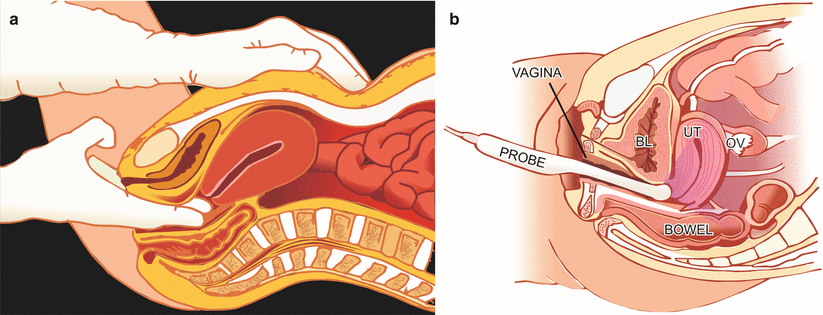
Today, with ultrasound, we can actually see pathology, and with a transvaginal ultrasound (TVS), one can not only see the pathology at close range, but also simultaneously touch the various structures that are seen, making it a dynamic and interactive examination (Fig. ). This is a distinct advantage of TVS over CT and MRI. Presently, ultrasound technology has advanced to such an extent that one can see on ultrasound almost as much as a pathologist can see on gross examination of a specimen (both the external surface and cut sections). Not only that, one can in addition see flow patterns within the mass which cannot be ascertained by gross examination of the specimen.
Fig 21 a Bimanual per vaginal examination which uses touch sensation to - photo 1
Fig. 2.1
( a ) Bimanual per vaginal examination which uses touch sensation to assess pathology of pelvic structures; ( b ) with TVS, which allows one to see pathology while touching the various structures simultaneously
Ideally a transabdominal scan (TAS) is done first with a full bladder, followed by a transvaginal scan (TVS) after having emptied the bladder. It is important that the technique is standardised, fixed and predetermined.
2.1 Transabdominal Scan
Filling the Bladder
For a good transabdominal scan, the bladder should be sufficiently filled, so as to push the bowels that lie in front of the pelvic organs towards the upper abdomen. Another advantage of a full bladder is that urine, being fluid, enhances the sound waves of the ultrasound beam, resulting in better visualisation. The bladder should not be over-distended because that would cause discomfort to the patient. In addition, a very full bladder increases the distance of the uterus and ovaries from the probe, decreasing resolution and resulting in suboptimal visualisation. A few points to be kept in mind here are:
  1. Very often one may be able to see sufficiently well on TAS even with a suboptimally filled bladder. Especially in cases that are to be followed by TVS, there is no need to insist that the bladder should be further filled.
  2. In emergency cases (like a case of a ruptured ectopic pregnancy), waiting for the bladder to fill may not be justified.
  3. In cases with a previous caesarean, the bladder may be adherent to the uterus at the site of the LSCS scar, and filling the bladder so as to visualise the upper uterus may not be possible. Instead, the more the patient fills her bladder, the more the lower uterine body and cervix get stretched, causing discomfort.
  4. With a very large and bulky uterus, a full bladder may not be able to overlie the entire uterus. Very often in these cases, the bulky uterus itself pushes the intestines out of the pelvis into the upper abdomen.
Next page
Light

Font size:

Reset

Interval:

Bookmark:

Make

Similar books «Ultrasound in Gynecology: an Atlas and Guide»

Look at similar books to Ultrasound in Gynecology: an Atlas and Guide. We have selected literature similar in name and meaning in the hope of providing readers with more options to find new, interesting, not yet read works.


Reviews about «Ultrasound in Gynecology: an Atlas and Guide»

Discussion, reviews of the book Ultrasound in Gynecology: an Atlas and Guide and just readers' own opinions. Leave your comments, write what you think about the work, its meaning or the main characters. Specify what exactly you liked and what you didn't like, and why you think so.